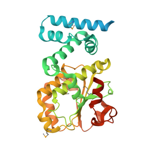
3HX3

Crystal structure of CRALBP mutant R234W


Experimental Data Snapshot

  • Method: X-RAY DIFFRACTION
  • Resolution: 1.69 Å
  • R-Value Free: 0.184 
  • R-Value Work: 0.167 
  • R-Value Observed: 0.169 

wwPDB Validation   3D Report Full Report


Ligand Structure Quality Assessment 


This is version 1.3 of the entry. See complete history


Literature

Bothnia dystrophy is caused by domino-like rearrangements in cellular retinaldehyde-binding protein mutant R234W.

He, X.Lobsiger, J.Stocker, A.

(2009) Proc Natl Acad Sci U S A 106: 18545-18550

  • DOI: https://doi.org/10.1073/pnas.0907454106
  • Primary Citation of Related Structures:  
    3HX3, 3HY5

  • PubMed Abstract: 

    Cellular retinaldehyde-binding protein (CRALBP) is essential for mammalian vision by routing 11-cis-retinoids for the conversion of photobleached opsin molecules into photosensitive visual pigments. The arginine-to-tryptophan missense mutation in position 234 (R234W) in the human gene RLBP1 encoding CRALBP compromises visual pigment regeneration and is associated with Bothnia dystrophy. Here we report the crystal structures of both wild-type human CRALBP and of its mutant R234W as binary complexes complemented with the endogenous ligand 11-cis-retinal, at 3.0 and 1.7 A resolution, respectively. Our structural model of wild-type CRALBP locates R234 to a positively charged cleft at a distance of 15 A from the hydrophobic core sequestering 11-cis-retinal. The R234W structural model reveals burial of W234 and loss of dianion-binding interactions within the cleft with physiological implications for membrane docking. The burial of W234 is accompanied by a cascade of side-chain flips that effect the intrusion of the side-chain of I238 into the ligand-binding cavity. As consequence of the intrusion, R234W displays 5-fold increased resistance to light-induced photoisomerization relative to wild-type CRALBP, indicating tighter binding to 11-cis-retinal. Overall, our results reveal an unanticipated domino-like structural transition causing Bothnia-type retinal dystrophy by the impaired release of 11-cis-retinal from R234W.


  • Organizational Affiliation

    Department of Chemistry and Biochemistry, University of Bern, 3012 Bern, Switzerland.


Macromolecules
Find similar proteins by:  (by identity cutoff)  |  3D Structure
Entity ID: 1
MoleculeChains Sequence LengthOrganismDetailsImage
Retinaldehyde-binding protein 1316Homo sapiensMutation(s): 1 
Gene Names: CRALBPRLBP1
UniProt & NIH Common Fund Data Resources
Find proteins for P12271 (Homo sapiens)
Explore P12271 
Go to UniProtKB:  P12271
PHAROS:  P12271
GTEx:  ENSG00000140522 
Entity Groups  
Sequence Clusters30% Identity50% Identity70% Identity90% Identity95% Identity100% Identity
UniProt GroupP12271
Sequence Annotations
Expand
  • Reference Sequence
Small Molecules
Ligands 1 Unique
IDChains Name / Formula / InChI Key2D Diagram3D Interactions
RET
Query on RET

Download Ideal Coordinates CCD File 
B [auth A]RETINAL
C20 H28 O
NCYCYZXNIZJOKI-OVSJKPMPSA-N
Modified Residues  1 Unique
IDChains TypeFormula2D DiagramParent
MSE
Query on MSE
A
L-PEPTIDE LINKINGC5 H11 N O2 SeMET
Binding Affinity Annotations 
IDSourceBinding Affinity
RET PDBBind:  3HX3 Kd: 10 (nM) from 1 assay(s)
Experimental Data & Validation

Experimental Data

  • Method: X-RAY DIFFRACTION
  • Resolution: 1.69 Å
  • R-Value Free: 0.184 
  • R-Value Work: 0.167 
  • R-Value Observed: 0.169 
  • Space Group: C 1 2 1
Unit Cell:
Length ( Å )Angle ( ˚ )
a = 87.93α = 90
b = 57.88β = 122.85
c = 75.15γ = 90
Software Package:
Software NamePurpose
XSCALEdata scaling
PHASERphasing
RESOLVEphasing
PHENIXrefinement
PDB_EXTRACTdata extraction
MOSFLMdata reduction
XDSdata reduction
XDSdata scaling

Structure Validation

View Full Validation Report


Ligand Structure Quality Assessment 


Entry History 

Deposition Data

Revision History  (Full details and data files)

  • Version 1.0: 2009-10-20
    Type: Initial release
  • Version 1.1: 2011-07-13
    Changes: Version format compliance
  • Version 1.2: 2021-10-13
    Changes: Database references, Derived calculations
  • Version 1.3: 2024-10-09
    Changes: Data collection, Structure summary