3HVP | pdb_00003hvp

CONSERVED FOLDING IN RETROVIRAL PROTEASES. CRYSTAL STRUCTURE OF A SYNTHETIC HIV-1 PROTEASE


Experimental Data Snapshot

  • Method: X-RAY DIFFRACTION
  • Resolution: 2.80 Å
  • R-Value Observed: 
    0.184 (Depositor) 

wwPDB Validation 3D Report Full Report

Validation slider image for 3HVP

This is version 1.4 of the entry. See complete history

Literature

Conserved folding in retroviral proteases: crystal structure of a synthetic HIV-1 protease.

Wlodawer, A.Miller, M.Jaskolski, M.Sathyanarayana, B.K.Baldwin, E.Weber, I.T.Selk, L.M.Clawson, L.Schneider, J.Kent, S.B.

(1989) Science 245: 616-621

  • DOI: https://doi.org/10.1126/science.2548279
  • Primary Citation Related Structures: 
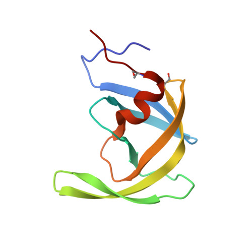
    3HVP

  • PubMed Abstract: 

    The rational design of drugs that can inhibit the action of viral proteases depends on obtaining accurate structures of these enzymes. The crystal structure of chemically synthesized HIV-1 protease has been determined at 2.8 angstrom resolution (R factor of 0.184) with the use of a model based on the Rous sarcoma virus protease structure. In this enzymatically active protein, the cysteines were replaced by alpha-amino-n-butyric acid, a nongenetically coded amino acid. This structure, in which all 99 amino acids were located, differs in several important details from that reported previously by others. The interface between the identical subunits forming the active protease dimer is composed of four well-ordered beta strands from both the amino and carboxyl termini and residues 86 to 94 have a helical conformation. The observed arrangement of the dimer interface suggests possible designs for dimerization inhibitors.


  • Organizational Affiliation
    • Crystallography Laboratory, NCI-Frederick Cancer Research Facility, MD 21701.

Macromolecule Content 

  • Total Structure Weight: 10.76 kDa 
  • Atom Count: 758 
  • Modeled Residue Count: 99 
  • Deposited Residue Count: 99 
  • Unique protein chains: 1

Macromolecules

Find similar proteins by:|  3D Structure
Entity ID: 1
MoleculeChains  Sequence LengthOrganismDetailsImage
UNLIGANDED HIV-1 PROTEASE99Human immunodeficiency virus 1Mutation(s): 0 
UniProt
Find proteins for P03369 (Human immunodeficiency virus type 1 group M subtype B (isolate ARV2/SF2))
Explore P03369 
Go to UniProtKB:  P03369
Entity Groups
Sequence Clusters30% Identity50% Identity70% Identity90% Identity95% Identity100% Identity
UniProt GroupP03369
Sequence Annotations
Expand
Reference Sequence

Small Molecules

Modified Residues  1 Unique
IDChains TypeFormula2D DiagramParent
ABA
Query on ABA
A
L-PEPTIDE LINKINGC4 H9 N O2ALA

Experimental Data & Validation

Experimental Data

  • Method: X-RAY DIFFRACTION
  • Resolution: 2.80 Å
  • R-Value Observed: 0.184 (Depositor) 
Space Group: P 41 21 2
Unit Cell:
Length ( Å )Angle ( ˚ )
a = 50.24α = 90
b = 50.24β = 90
c = 106.56γ = 90
Software Package:
Software NamePurpose
PROFFTrefinement

Structure Validation

View Full Validation Report


Entry History 

Deposition Data

Revision History  (Full details and data files)

  • Version 1.0: 1989-10-15
    Type: Initial release
  • Version 1.1: 2008-03-03
    Changes: Version format compliance
  • Version 1.2: 2011-07-13
    Changes: Derived calculations, Version format compliance
  • Version 1.3: 2017-11-29
    Changes: Derived calculations, Other
  • Version 1.4: 2025-03-26
    Changes: Data collection, Database references, Derived calculations, Structure summary