3HVI | pdb_00003hvi

Rat catechol O-methyltransferase in complex with a catechol-type, N6-ethyladenine-containing bisubstrate inhibitor


Experimental Data Snapshot

  • Method: X-RAY DIFFRACTION
  • Resolution: 1.20 Å
  • R-Value Free: 
    0.161 (Depositor), 0.159 (DCC) 
  • R-Value Work: 
    0.131 (Depositor), 0.129 (DCC) 
  • R-Value Observed: 
    0.132 (Depositor) 

wwPDB Validation 3D Report Full Report

Validation slider image for 3HVI

Ligand Structure Quality Assessment 


This is version 1.4 of the entry. See complete history

Literature

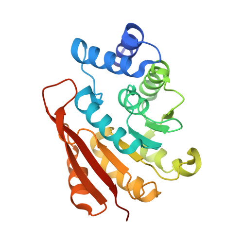
Molecular recognition at the active site of catechol-o-methyltransferase: energetically favorable replacement of a water molecule imported by a bisubstrate inhibitor.

Ellermann, M.Jakob-Roetne, R.Lerner, C.Borroni, E.Schlatter, D.Roth, D.Ehler, A.Rudolph, M.G.Diederich, F.

(2009) Angew Chem Int Ed Engl 48: 9092-9096

Macromolecule Content 

  • Total Structure Weight: 25.56 kDa 
  • Atom Count: 2,211 
  • Modeled Residue Count: 213 
  • Deposited Residue Count: 221 
  • Unique protein chains: 1

Macromolecules

Find similar proteins by:|  3D Structure
Entity ID: 1
MoleculeChains  Sequence LengthOrganismDetailsImage
Catechol O-methyltransferase221Rattus norvegicusMutation(s): 0 
EC: 2.1.1.6
UniProt
Find proteins for P22734 (Rattus norvegicus)
Explore P22734 
Go to UniProtKB:  P22734
Entity Groups
Sequence Clusters30% Identity50% Identity70% Identity90% Identity95% Identity100% Identity
UniProt GroupP22734
Sequence Annotations
Expand
Reference Sequence

Small Molecules

Ligands 5 Unique
IDChains Name / Formula / InChI Key2D Diagram3D Interactions
619

Query on 619


Download:Ideal Coordinates CCD File
E [auth A]N-[(E)-3-[(2R,3S,4R,5R)-5-(6-ethylaminopurin-9-yl)-3,4-dihydroxy-oxolan-2-yl]prop-2-enyl]-5-(4-fluorophenyl)-2,3-dihydroxy-benzamide
C27 H27 F N6 O6
NOCNCHHJBSJZFZ-CJLJSIFTSA-N
D1D

Query on D1D


Download:Ideal Coordinates CCD File
F [auth A](4S,5S)-1,2-DITHIANE-4,5-DIOL
C4 H8 O2 S2
YPGMOWHXEQDBBV-QWWZWVQMSA-N
CL

Query on CL


Download:Ideal Coordinates CCD File
D [auth A]CHLORIDE ION
Cl
VEXZGXHMUGYJMC-UHFFFAOYSA-M
MG

Query on MG


Download:Ideal Coordinates CCD File
B [auth A]MAGNESIUM ION
Mg
JLVVSXFLKOJNIY-UHFFFAOYSA-N
NA

Query on NA


Download:Ideal Coordinates CCD File
C [auth A]SODIUM ION
Na
FKNQFGJONOIPTF-UHFFFAOYSA-N

Experimental Data & Validation

Experimental Data

  • Method: X-RAY DIFFRACTION
  • Resolution: 1.20 Å
  • R-Value Free:  0.161 (Depositor), 0.159 (DCC) 
  • R-Value Work:  0.131 (Depositor), 0.129 (DCC) 
  • R-Value Observed: 0.132 (Depositor) 
Space Group: P 21 21 21
Unit Cell:
Length ( Å )Angle ( ˚ )
a = 49.857α = 90
b = 54.706β = 90
c = 80.617γ = 90
Software Package:
Software NamePurpose
COMOphasing
REFMACrefinement
XDSdata reduction
SADABSdata scaling

Structure Validation

View Full Validation Report


Ligand Structure Quality Assessment 


Entry History 

Deposition Data

Revision History  (Full details and data files)

  • Version 1.0: 2009-10-13
    Type: Initial release
  • Version 1.1: 2011-07-13
    Changes: Version format compliance
  • Version 1.2: 2012-08-08
    Changes: Database references, Structure summary
  • Version 1.3: 2017-11-01
    Changes: Refinement description
  • Version 1.4: 2024-03-20
    Changes: Data collection, Database references, Derived calculations