3HSS | pdb_00003hss

A higher resolution structure of Rv0554 from Mycobacterium tuberculosis complexed with malonic acid


Experimental Data Snapshot

  • Method: X-RAY DIFFRACTION
  • Resolution: 1.90 Å
  • R-Value Free: 
    0.219 (Depositor), 0.212 (DCC) 
  • R-Value Work: 
    0.193 (Depositor), 0.184 (DCC) 
  • R-Value Observed: 
    0.194 (Depositor) 

wwPDB Validation 3D Report Full Report

Validation slider image for 3HSS

This is version 1.4 of the entry. See complete history

Literature

Structural and functional analysis of Rv0554 from Mycobacterium tuberculosis: testing a putative role in menaquinone biosynthesis.

Johnston, J.M.Jiang, M.Guo, Z.Baker, E.N.

(2010) Acta Crystallogr D Biol Crystallogr 66: 909-917

  • DOI: https://doi.org/10.1107/S0907444910025771
  • Primary Citation Related Structures: 
    3E3A, 3HSS, 3HYS

  • PubMed Abstract: 

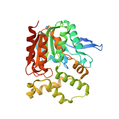
    Mycobacterium tuberculosis, the cause of tuberculosis, is a devastating human pathogen against which new drugs are urgently needed. Enzymes from the biosynthetic pathway for menaquinone are considered to be valid drug targets. The protein encoded by the open reading frame Rv0554 has been expressed, purified and subjected to structural and functional analysis to test for a putative role in menaquinone biosynthesis. The crystal structure of Rv0554 has been solved and refined in two different space groups at 2.35 and 1.9 A resolution. The protein is dimeric, with an alpha/beta-hydrolase monomer fold. In each monomer, a large cavity adjacent to the catalytic triad is enclosed by a helical lid. Dimerization is mediated by the lid regions. Small-molecule additives used in crystallization bind in the active site, but no binding of ligands related to menaquinone biosynthesis could be detected and functional assays failed to support possible roles in menaquinone biosynthesis.


  • Organizational Affiliation
    • School of Biological Sciences, University of Auckland, Private Bag 92019, Auckland, New Zealand.

Macromolecule Content 

  • Total Structure Weight: 65.42 kDa 
  • Atom Count: 4,705 
  • Modeled Residue Count: 535 
  • Deposited Residue Count: 586 
  • Unique protein chains: 1

Macromolecules

Find similar proteins by:|  3D Structure
Entity ID: 1
MoleculeChains  Sequence LengthOrganismDetailsImage
putative Bromoperoxidase
A, B
293Mycobacterium tuberculosisMutation(s): 0 
Gene Names: bpoCMT0580Rv0554
EC: 1.11.1
UniProt
Find proteins for P9WNH1 (Mycobacterium tuberculosis (strain ATCC 25618 / H37Rv))
Explore P9WNH1 
Go to UniProtKB:  P9WNH1
Entity Groups
Sequence Clusters30% Identity50% Identity70% Identity90% Identity95% Identity100% Identity
UniProt GroupP9WNH1
Sequence Annotations
Expand
Reference Sequence

Small Molecules

Ligands 5 Unique
IDChains Name / Formula / InChI Key2D Diagram3D Interactions
TRS

Query on TRS


Download:Ideal Coordinates CCD File
C [auth A]2-AMINO-2-HYDROXYMETHYL-PROPANE-1,3-DIOL
C4 H12 N O3
LENZDBCJOHFCAS-UHFFFAOYSA-O
MLA

Query on MLA


Download:Ideal Coordinates CCD File
E [auth A],
L [auth B],
O [auth B]
MALONIC ACID
C3 H4 O4
OFOBLEOULBTSOW-UHFFFAOYSA-N
EDO

Query on EDO


Download:Ideal Coordinates CCD File
D [auth A]
F [auth A]
G [auth A]
H [auth A]
K [auth A]
D [auth A],
F [auth A],
G [auth A],
H [auth A],
K [auth A],
M [auth B]
1,2-ETHANEDIOL
C2 H6 O2
LYCAIKOWRPUZTN-UHFFFAOYSA-N
ACT

Query on ACT


Download:Ideal Coordinates CCD File
J [auth A]ACETATE ION
C2 H3 O2
QTBSBXVTEAMEQO-UHFFFAOYSA-M
NA

Query on NA


Download:Ideal Coordinates CCD File
I [auth A],
N [auth B]
SODIUM ION
Na
FKNQFGJONOIPTF-UHFFFAOYSA-N

Experimental Data & Validation

Experimental Data

  • Method: X-RAY DIFFRACTION
  • Resolution: 1.90 Å
  • R-Value Free:  0.219 (Depositor), 0.212 (DCC) 
  • R-Value Work:  0.193 (Depositor), 0.184 (DCC) 
  • R-Value Observed: 0.194 (Depositor) 
Space Group: P 41 21 2
Unit Cell:
Length ( Å )Angle ( ˚ )
a = 100.454α = 90
b = 100.454β = 90
c = 135.162γ = 90
Software Package:
Software NamePurpose
DENZOdata reduction
SCALEPACKdata scaling
MOLREPphasing
REFMACrefinement
PDB_EXTRACTdata extraction
HKL-2000data collection
HKL-2000data reduction
HKL-2000data scaling

Structure Validation

View Full Validation Report


Entry History 

Deposition Data

Revision History  (Full details and data files)

  • Version 1.0: 2010-05-26
    Type: Initial release
  • Version 1.1: 2011-07-13
    Changes: Version format compliance
  • Version 1.2: 2015-04-29
    Changes: Non-polymer description
  • Version 1.3: 2017-11-01
    Changes: Advisory, Refinement description
  • Version 1.4: 2024-02-21
    Changes: Advisory, Data collection, Database references, Derived calculations