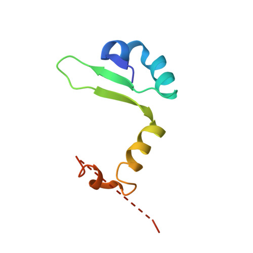
3HRY

Crystal structure of PHD in a trigonal space group and partially disordered


Experimental Data Snapshot

  • Method: X-RAY DIFFRACTION
  • Resolution: 2.25 Å
  • R-Value Free: 0.256 
  • R-Value Work: 0.192 
  • R-Value Observed: 0.195 

Starting Model: experimental
View more details

wwPDB Validation   3D Report Full Report


This is version 1.2 of the entry. See complete history


Literature

Allostery and intrinsic disorder mediate transcription regulation by conditional cooperativity.

Garcia-Pino, A.Balasubramanian, S.Wyns, L.Gazit, E.De Greve, H.Magnuson, R.D.Charlier, D.van Nuland, N.A.Loris, R.

(2010) Cell 142: 101-111

  • DOI: https://doi.org/10.1016/j.cell.2010.05.039
  • Primary Citation of Related Structures:  
    3HRY, 3HS2, 3K33

  • PubMed Abstract: 

    Regulation of the phd/doc toxin-antitoxin operon involves the toxin Doc as co- or derepressor depending on the ratio between Phd and Doc, a phenomenon known as conditional cooperativity. The mechanism underlying this observed behavior is not understood. Here we show that monomeric Doc engages two Phd dimers on two unrelated binding sites. The binding of Doc to the intrinsically disordered C-terminal domain of Phd structures its N-terminal DNA-binding domain, illustrating allosteric coupling between highly disordered and highly unstable domains. This allosteric effect also couples Doc neutralization to the conditional regulation of transcription. In this way, higher levels of Doc tighten repression up to a point where the accumulation of toxin triggers the production of Phd to counteract its action. Our experiments provide the basis for understanding the mechanism of conditional cooperative regulation of transcription typical of toxin-antitoxin modules. This model may be applicable for the regulation of other biological systems.


  • Organizational Affiliation

    Structural Biology Brussels, Vrije Universiteit Brussels, Pleinlaan 2, B-1050 Brussels, Belgium. agarciap@vub.ac.be


Macromolecules
Find similar proteins by:  (by identity cutoff)  |  3D Structure
Entity ID: 1
MoleculeChains Sequence LengthOrganismDetailsImage
Prevent host death protein
A, B, C
73Escherichia coliMutation(s): 0 
Gene Names: phdPhdIPF_68
UniProt
Find proteins for Q06253 (Escherichia phage P1)
Explore Q06253 
Go to UniProtKB:  Q06253
Entity Groups  
Sequence Clusters30% Identity50% Identity70% Identity90% Identity95% Identity100% Identity
UniProt GroupQ06253
Sequence Annotations
Expand
  • Reference Sequence
Experimental Data & Validation

Experimental Data

  • Method: X-RAY DIFFRACTION
  • Resolution: 2.25 Å
  • R-Value Free: 0.256 
  • R-Value Work: 0.192 
  • R-Value Observed: 0.195 
  • Space Group: P 31 2 1
Unit Cell:
Length ( Å )Angle ( ˚ )
a = 71.951α = 90
b = 71.951β = 90
c = 68.007γ = 120
Software Package:
Software NamePurpose
MSdata collection
PHASERphasing
PHENIXrefinement
DENZOdata reduction
SCALEPACKdata scaling

Structure Validation

View Full Validation Report


Entry History 

Deposition Data

Revision History  (Full details and data files)

  • Version 1.0: 2010-06-23
    Type: Initial release
  • Version 1.1: 2011-07-13
    Changes: Version format compliance
  • Version 1.2: 2023-09-06
    Changes: Data collection, Database references, Refinement description