3HHF | pdb_00003hhf

Structure of CrgA regulatory domain, a LysR-type transcriptional regulator from Neisseria meningitidis.


Experimental Data Snapshot

  • Method: X-RAY DIFFRACTION
  • Resolution: 2.30 Å
  • R-Value Free: 
    0.258 (Depositor), 0.254 (DCC) 
  • R-Value Work: 
    0.201 (Depositor), 0.204 (DCC) 
  • R-Value Observed: 
    0.204 (Depositor) 

wwPDB Validation 3D Report Full Report

Validation slider image for 3HHF

This is version 1.2 of the entry. See complete history

Literature

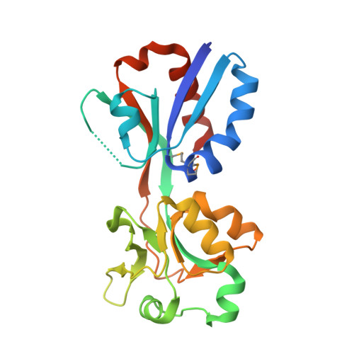
The structure of CrgA from Neisseria meningitidis reveals a new octameric assembly state for LysR transcriptional regulators

Sainsbury, S.Lane, L.A.Ren, J.Gilbert, R.J.Saunders, N.J.Robinson, C.V.Stuart, D.I.Owens, R.J.

(2009) Nucleic Acids Res 37: 4545-4558

  • DOI: https://doi.org/10.1093/nar/gkp445
  • Primary Citation Related Structures: 
    3HHF, 3HHG

  • PubMed Abstract: 

    LysR-type transcriptional regulators (LTTRs) form the largest family of bacterial regulators acting as both auto-repressors and activators of target promoters, controlling operons involved in a wide variety of cellular processes. The LTTR, CrgA, from the human pathogen Neisseria meningitidis, is upregulated during bacterial-host cell contact. Here, we report the crystal structures of both regulatory domain and full-length CrgA, the first of a novel subclass of LTTRs that form octameric rings. Non-denaturing mass spectrometry analysis and analytical ultracentrifugation established that the octameric form of CrgA is the predominant species in solution in both the presence and absence of an oligonucleotide encompassing the CrgA-binding sequence. Furthermore, analysis of the isolated CrgA-DNA complex by mass spectrometry showed stabilization of a double octamer species upon DNA binding. Based on the observed structure and the mass spectrometry findings, a model is proposed in which a hexadecameric array of two CrgA oligomers binds to its DNA target site.


  • Organizational Affiliation
    • The Oxford Protein Production Facility and Division of Structural Biology, Henry Wellcome Building for Genomic Medicine, University of Oxford, Roosevelt Drive, Oxford OX3 7BN, UK.

Macromolecule Content 

  • Total Structure Weight: 46.95 kDa 
  • Atom Count: 3,344 
  • Modeled Residue Count: 406 
  • Deposited Residue Count: 426 
  • Unique protein chains: 1

Macromolecules

Find similar proteins by:|  3D Structure
Entity ID: 1
MoleculeChains  Sequence LengthOrganismDetailsImage
Transcriptional regulator, LysR familyA [auth B],
B [auth A]
213Neisseria meningitidis serogroup BMutation(s): 0 
Gene Names: CrgANMB1856
UniProt
Find proteins for Q9JXW7 (Neisseria meningitidis serogroup B (strain ATCC BAA-335 / MC58))
Explore Q9JXW7 
Go to UniProtKB:  Q9JXW7
Entity Groups
Sequence Clusters30% Identity50% Identity70% Identity90% Identity95% Identity100% Identity
UniProt GroupQ9JXW7
Sequence Annotations
Expand
Reference Sequence

Small Molecules

Modified Residues  1 Unique
IDChains TypeFormula2D DiagramParent
MSE
Query on MSE
A [auth B],
B [auth A]
L-PEPTIDE LINKINGC5 H11 N O2 SeMET

Experimental Data & Validation

Experimental Data

  • Method: X-RAY DIFFRACTION
  • Resolution: 2.30 Å
  • R-Value Free:  0.258 (Depositor), 0.254 (DCC) 
  • R-Value Work:  0.201 (Depositor), 0.204 (DCC) 
  • R-Value Observed: 0.204 (Depositor) 
Space Group: P 1 21 1
Unit Cell:
Length ( Å )Angle ( ˚ )
a = 51.6α = 90
b = 65.6β = 105
c = 63.5γ = 90
Software Package:
Software NamePurpose
HKL-2000data collection
SOLVEphasing
REFMACrefinement
HKL-2000data reduction
SCALEPACKdata scaling

Structure Validation

View Full Validation Report


Entry History 

Deposition Data

Revision History  (Full details and data files)

  • Version 1.0: 2009-07-07
    Type: Initial release
  • Version 1.1: 2011-07-13
    Changes: Advisory, Version format compliance
  • Version 1.2: 2024-11-27
    Changes: Data collection, Database references, Derived calculations, Refinement description, Structure summary