


Structural basis for anionic ligand recognition by multidrug binding proteins: crystal structures of CmeR-bile acid complexes
Routh, M.D., Yang, F., Su, C.-C., Zhang, Q., Yu, E.W.To be published.
Experimental Data Snapshot
Entity ID: 1 | |||||
|---|---|---|---|---|---|
| Molecule | Chains | Sequence Length | Organism | Details | Image |
| CmeR | 216 | Campylobacter jejuni | Mutation(s): 0 | 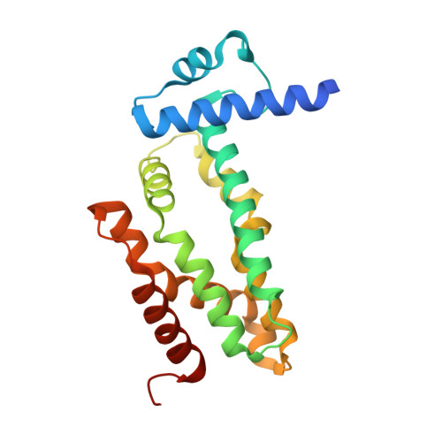 | |
UniProt | |||||
Entity Groups | |||||
| Sequence Clusters | 30% Identity50% Identity70% Identity90% Identity95% Identity100% Identity | ||||
| UniProt Group | Q7B8P6 | ||||
Sequence AnnotationsExpand | |||||
Reference Sequence | |||||
| Ligands 1 Unique | |||||
|---|---|---|---|---|---|
| ID | Chains | Name / Formula / InChI Key | 2D Diagram | 3D Interactions | |
| TCH Download:Ideal Coordinates CCD File | B [auth A] | TAUROCHOLIC ACID C26 H45 N O7 S WBWWGRHZICKQGZ-HZAMXZRMSA-N |  | ||
| Length ( Å ) | Angle ( ˚ ) |
|---|---|
| a = 93.69 | α = 90 |
| b = 37.488 | β = 90 |
| c = 57.733 | γ = 90 |
| Software Name | Purpose |
|---|---|
| DENZO | data reduction |
| SCALEPACK | data scaling |
| PHASER | phasing |
| PHENIX | refinement |
| PDB_EXTRACT | data extraction |
| ADSC | data collection |


