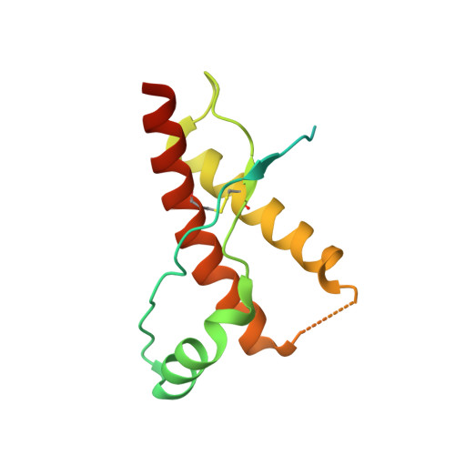
3HES

Human prion protein variant F198S with M129


Experimental Data Snapshot

  • Method: X-RAY DIFFRACTION
  • Resolution: 2.00 Å
  • R-Value Free: 0.249 
  • R-Value Work: 0.216 
  • R-Value Observed: 0.217 

wwPDB Validation   3D Report Full Report


This is version 1.4 of the entry. See complete history


Literature

Conformational diversity in prion protein variants influences intermolecular beta-sheet formation.

Lee, S.Antony, L.Hartmann, R.Knaus, K.J.Surewicz, K.Surewicz, W.K.Yee, V.C.

(2010) EMBO J 29: 251-262

  • DOI: https://doi.org/10.1038/emboj.2009.333
  • Primary Citation of Related Structures:  
    3HAF, 3HAK, 3HEQ, 3HER, 3HES, 3HJ5, 3HJX

  • PubMed Abstract: 

    A conformational transition of normal cellular prion protein (PrP(C)) to its pathogenic form (PrP(Sc)) is believed to be a central event in the transmission of the devastating neurological diseases known as spongiform encephalopathies. The common methionine/valine polymorphism at residue 129 in the PrP influences disease susceptibility and phenotype. We report here seven crystal structures of human PrP variants: three of wild-type (WT) PrP containing V129, and four of the familial variants D178N and F198S, containing either M129 or V129. Comparison of these structures with each other and with previously published WT PrP structures containing M129 revealed that only WT PrPs were found to crystallize as domain-swapped dimers or closed monomers; the four mutant PrPs crystallized as non-swapped dimers. Three of the four mutant PrPs aligned to form intermolecular beta-sheets. Several regions of structural variability were identified, and analysis of their conformations provides an explanation for the structural features, which can influence the formation and conformation of intermolecular beta-sheets involving the M/V129 polymorphic residue.


  • Organizational Affiliation

    Department of Biochemistry, Case Western Reserve University, Cleveland, OH 44106, USA.


Macromolecules
Find similar proteins by:  (by identity cutoff)  |  3D Structure
Entity ID: 1
MoleculeChains Sequence LengthOrganismDetailsImage
Major prion protein
A, B
142Homo sapiensMutation(s): 1 
Gene Names: PRNPPRIPPRP
UniProt & NIH Common Fund Data Resources
Find proteins for P04156 (Homo sapiens)
Explore P04156 
Go to UniProtKB:  P04156
PHAROS:  P04156
GTEx:  ENSG00000171867 
Entity Groups  
Sequence Clusters30% Identity50% Identity70% Identity90% Identity95% Identity100% Identity
UniProt GroupP04156
Sequence Annotations
Expand
  • Reference Sequence
Experimental Data & Validation

Experimental Data

  • Method: X-RAY DIFFRACTION
  • Resolution: 2.00 Å
  • R-Value Free: 0.249 
  • R-Value Work: 0.216 
  • R-Value Observed: 0.217 
  • Space Group: P 43 21 2
Unit Cell:
Length ( Å )Angle ( ˚ )
a = 57.017α = 90
b = 57.017β = 90
c = 167.364γ = 90
Software Package:
Software NamePurpose
DENZOdata reduction
SCALEPACKdata scaling
REFMACrefinement
PDB_EXTRACTdata extraction
HKL-2000data collection
HKL-2000data reduction
SOLVEphasing

Structure Validation

View Full Validation Report


Entry History 

Deposition Data

Revision History  (Full details and data files)

  • Version 1.0: 2010-01-12
    Type: Initial release
  • Version 1.1: 2011-07-13
    Changes: Version format compliance
  • Version 1.2: 2017-11-01
    Changes: Refinement description
  • Version 1.3: 2021-10-13
    Changes: Database references, Derived calculations
  • Version 1.4: 2024-10-30
    Changes: Data collection, Structure summary