


Domain swapping promoted by a single mutation that introduces an ionizable group into the hydrophobic core of a protein
Khangulov, V.S., Schlessman, J.L., Heroux, A., Garcia-Moreno, E.B.To be published.
Experimental Data Snapshot
Entity ID: 1 | |||||
|---|---|---|---|---|---|
| Molecule | Chains | Sequence Length | Organism | Details | Image |
| Thermonuclease | A, B [auth D], C [auth B], D [auth C] | 143 | Staphylococcus aureus | Mutation(s): 6 Gene Names: nuc EC: 3.1.31.1 | 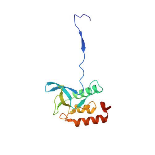 |
UniProt | |||||
Entity Groups | |||||
| Sequence Clusters | 30% Identity50% Identity70% Identity90% Identity95% Identity100% Identity | ||||
| UniProt Group | P00644 | ||||
Sequence AnnotationsExpand | |||||
Reference Sequence | |||||
| Ligands 4 Unique | |||||
|---|---|---|---|---|---|
| ID | Chains | Name / Formula / InChI Key | 2D Diagram | 3D Interactions | |
| THP Download:Ideal Coordinates CCD File | E [auth A], H [auth D], I [auth B], K [auth C] | THYMIDINE-3',5'-DIPHOSPHATE C10 H16 N2 O11 P2 CSNCBOPUCJOHLS-XLPZGREQSA-N |  | ||
| MRD Download:Ideal Coordinates CCD File | J [auth B] | (4R)-2-METHYLPENTANE-2,4-DIOL C6 H14 O2 SVTBMSDMJJWYQN-RXMQYKEDSA-N |  | ||
| MPD Download:Ideal Coordinates CCD File | F [auth A] | (4S)-2-METHYL-2,4-PENTANEDIOL C6 H14 O2 SVTBMSDMJJWYQN-YFKPBYRVSA-N |  | ||
| GOL Download:Ideal Coordinates CCD File | G [auth A], L [auth C] | GLYCEROL C3 H8 O3 PEDCQBHIVMGVHV-UHFFFAOYSA-N |  | ||
| Length ( Å ) | Angle ( ˚ ) |
|---|---|
| a = 67.598 | α = 90 |
| b = 74.42 | β = 90.22 |
| c = 67.975 | γ = 90 |
| Software Name | Purpose |
|---|---|
| DENZO | data reduction |
| SCALEPACK | data scaling |
| PHASER | phasing |
| REFMAC | refinement |
| PDB_EXTRACT | data extraction |
| CBASS | data collection |
| HKL-2000 | data scaling |


