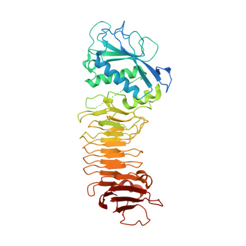
3HDA

PrtC methionine mutants: M226A_DESY


Experimental Data Snapshot

  • Method: X-RAY DIFFRACTION
  • Resolution: 2.13 Å
  • R-Value Free: 0.207 
  • R-Value Work: 0.182 
  • R-Value Observed: 0.183 

Starting Model: experimental
View more details

wwPDB Validation   3D Report Full Report


This is version 1.6 of the entry. See complete history


Literature

Metzincin's canonical methionine is responsible for the structural integrity of the zinc-binding site

Oberholzer, A.E.Bumann, M.Hege, T.Russo, S.Baumann, U.

(2009) Biol Chem 390: 875-881

  • DOI: https://doi.org/10.1515/BC.2009.100
  • Primary Citation of Related Structures:  
    3HB2, 3HBU, 3HBV, 3HDA

  • PubMed Abstract: 

    The metzincins constitute a subclan of metalloproteases possessing a HEXXHXXGXXH/D zinc-binding consensus sequence where the three histidines are zinc ligands and the glutamic acid is the catalytic base. A completely conserved methionine is located downstream of this motif. Families of the metzincin clan comprise, besides others, astacins, adamalysins proteases, matrix metallo-proteases, and serralysins. The latter are extracellular 50 kDa proteases secreted by Gram-negative bacteria via a type I secretion system. While there is a large body of structural and biochemical information available, the function of the conserved methionine has not been convincingly clarified yet. Here, we present the crystal structures of a number of mutants of the serralysin member protease C with the conserved methionine being replaced by Ile, Ala, and His. Together with our former report on the leucine and cysteine mutants, we demonstrate here that replacement of the methionine side chain results in an increasing distortion of the zinc-binding geometry, especially pronounced in the chi(2) angles of the first and third histidine of the consensus sequence. This is correlated with an increasing loss of proteolytic activity and a sharp increase of flexibility of large segments of the polypeptide chain.


  • Organizational Affiliation

    Department of Chemistry and Biochemistry, University of Bern, Bern, Switzerland.


Macromolecules
Find similar proteins by:  (by identity cutoff)  |  3D Structure
Entity ID: 1
MoleculeChains Sequence LengthOrganismDetailsImage
Secreted protease CA [auth P]462Dickeya chrysanthemiMutation(s): 1 
Gene Names: prtC
EC: 3.4.24 (PDB Primary Data), 3.4.24.40 (UniProt)
UniProt
Find proteins for P16317 (Dickeya chrysanthemi)
Explore P16317 
Go to UniProtKB:  P16317
Entity Groups  
Sequence Clusters30% Identity50% Identity70% Identity90% Identity95% Identity100% Identity
UniProt GroupP16317
Sequence Annotations
Expand
  • Reference Sequence

Find similar proteins by:  Sequence   |   3D Structure  

Entity ID: 2
MoleculeChains Sequence LengthOrganismDetailsImage
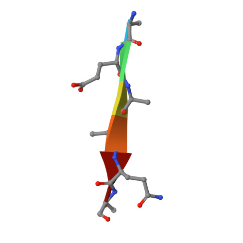
Uncharacterized peptideB [auth Z]6synthetic constructMutation(s): 0 
Sequence Annotations
Expand
  • Reference Sequence
Small Molecules
Ligands 2 Unique
IDChains Name / Formula / InChI Key2D Diagram3D Interactions
CA
Query on CA

Download Ideal Coordinates CCD File 
C [auth P]
D [auth P]
E [auth P]
F [auth P]
G [auth P]
C [auth P],
D [auth P],
E [auth P],
F [auth P],
G [auth P],
H [auth P],
I [auth P]
CALCIUM ION
Ca
BHPQYMZQTOCNFJ-UHFFFAOYSA-N
CL
Query on CL

Download Ideal Coordinates CCD File 
J [auth P],
K [auth P]
CHLORIDE ION
Cl
VEXZGXHMUGYJMC-UHFFFAOYSA-M
Experimental Data & Validation

Experimental Data

  • Method: X-RAY DIFFRACTION
  • Resolution: 2.13 Å
  • R-Value Free: 0.207 
  • R-Value Work: 0.182 
  • R-Value Observed: 0.183 
  • Space Group: P 31 2 1
Unit Cell:
Length ( Å )Angle ( ˚ )
a = 101.858α = 90
b = 101.858β = 90
c = 122.521γ = 120
Software Package:
Software NamePurpose
PHENIXrefinement
SCALEPACKdata scaling
CNSrefinement
DENZOdata reduction
PDB_EXTRACTdata extraction
HKL-2000data collection
CNSphasing

Structure Validation

View Full Validation Report


Entry History 

Deposition Data

Revision History  (Full details and data files)

  • Version 1.0: 2009-06-30
    Type: Initial release
  • Version 1.1: 2011-07-13
    Changes: Version format compliance
  • Version 1.2: 2016-02-17
    Changes: Refinement description
  • Version 1.3: 2017-11-01
    Changes: Refinement description
  • Version 1.4: 2019-07-24
    Changes: Data collection, Refinement description, Source and taxonomy
  • Version 1.5: 2021-10-13
    Changes: Database references, Derived calculations
  • Version 1.6: 2023-09-06
    Changes: Data collection, Refinement description