3HD6 | pdb_00003hd6

Crystal Structure of the Human Rhesus Glycoprotein RhCG


Experimental Data Snapshot

  • Method: X-RAY DIFFRACTION
  • Resolution: 2.10 Å
  • R-Value Free: 
    0.195 (Depositor), 0.190 (DCC) 
  • R-Value Work: 
    0.168 (Depositor) 
  • R-Value Observed: 
    0.170 (Depositor) 

Starting Model: experimental
View more details

wwPDB Validation 3D Report Full Report

Validation slider image for 3HD6

Ligand Structure Quality Assessment 


This is version 1.5 of the entry. See complete history

Literature

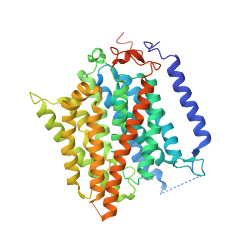
Function of human Rh based on structure of RhCG at 2.1 A.

Gruswitz, F.Chaudhary, S.Ho, J.D.Schlessinger, A.Pezeshki, B.Ho, C.M.Sali, A.Westhoff, C.M.Stroud, R.M.

(2010) Proc Natl Acad Sci U S A 107: 9638-9643

  • DOI: https://doi.org/10.1073/pnas.1003587107
  • Primary Citation Related Structures: 
    3HD6

  • PubMed Abstract: 

    In humans, NH(3) transport across cell membranes is facilitated by the Rh (rhesus) family of proteins. Human Rh C glycoprotein (RhCG) forms a trimeric complex that plays an essential role in ammonia excretion and renal pH regulation. The X-ray crystallographic structure of human RhCG, determined at 2.1 A resolution, reveals the mechanism of ammonia transport. Each monomer contains 12 transmembrane helices, one more than in the bacterial homologs. Reconstituted into proteoliposomes, RhCG conducts NH(3) to raise internal pH. Models of the erythrocyte Rh complex based on our RhCG structure suggest that the erythrocytic Rh complex is composed of stochastically assembled heterotrimers of RhAG, RhD, and RhCE.


  • Organizational Affiliation
    • Department of Biochemistry and Biophysics, S412C Genentech Hall, Center for the Structure of Membrane Proteins, and Membrane Protein Expression Center, University of California, San Francisco, CA 94158, USA.

Macromolecule Content 

  • Total Structure Weight: 54.47 kDa 
  • Atom Count: 3,299 
  • Modeled Residue Count: 403 
  • Deposited Residue Count: 490 
  • Unique protein chains: 1

Macromolecules

Find similar proteins by:|  3D Structure
Entity ID: 1
MoleculeChains  Sequence LengthOrganismDetailsImage
Ammonium transporter Rh type C490Homo sapiensMutation(s): 0 
Gene Names: C15orf6CDRC2PDRC2RHCGRHGK
Membrane Entity: Yes 
UniProt & NIH Common Fund Data Resources
Find proteins for Q9UBD6 (Homo sapiens)
Explore Q9UBD6 
Go to UniProtKB:  Q9UBD6
PHAROS:  Q9UBD6
GTEx:  ENSG00000140519 
Entity Groups
Sequence Clusters30% Identity50% Identity70% Identity90% Identity95% Identity100% Identity
UniProt GroupQ9UBD6
Sequence Annotations
Expand
Reference Sequence

Small Molecules

Ligands 1 Unique
IDChains Name / Formula / InChI Key2D Diagram3D Interactions
BOG

Query on BOG


Download:Ideal Coordinates CCD File
B [auth A]octyl beta-D-glucopyranoside
C14 H28 O6
HEGSGKPQLMEBJL-RKQHYHRCSA-N

Experimental Data & Validation

Experimental Data

  • Method: X-RAY DIFFRACTION
  • Resolution: 2.10 Å
  • R-Value Free:  0.195 (Depositor), 0.190 (DCC) 
  • R-Value Work:  0.168 (Depositor) 
  • R-Value Observed: 0.170 (Depositor) 
Space Group: P 63
Unit Cell:
Length ( Å )Angle ( ˚ )
a = 99.83α = 90
b = 99.83β = 90
c = 101.076γ = 120
Software Package:
Software NamePurpose
ADSCdata collection
PHASERphasing
REFMACrefinement
HKL-2000data reduction
HKL-2000data scaling

Structure Validation

View Full Validation Report


Ligand Structure Quality Assessment 


Entry History 

Deposition Data

Revision History  (Full details and data files)

  • Version 1.0: 2009-09-01
    Type: Initial release
  • Version 1.1: 2011-07-13
    Changes: Advisory, Version format compliance
  • Version 1.2: 2018-01-24
    Changes: Structure summary
  • Version 1.3: 2020-07-29
    Type: Remediation
    Reason: Carbohydrate remediation
    Changes: Data collection, Derived calculations, Structure summary
  • Version 1.4: 2021-03-31
    Changes: Source and taxonomy, Structure summary
  • Version 1.5: 2023-09-06
    Changes: Data collection, Database references, Refinement description