3HB6 | pdb_00003hb6

Inactive mutant H54F of Proteus mirabilis catalase


Experimental Data Snapshot

  • Method: X-RAY DIFFRACTION
  • Resolution: 2.30 Å
  • R-Value Free: 
    0.236 (Depositor), 0.228 (DCC) 
  • R-Value Work: 
    0.203 (Depositor), 0.198 (DCC) 
  • R-Value Observed: 
    0.203 (Depositor) 

Starting Model: experimental
View more details

wwPDB Validation 3D Report Full Report

Validation slider image for 3HB6

Ligand Structure Quality Assessment 


This is version 1.3 of the entry. See complete history

Literature

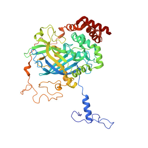
Verdoheme formation in Proteus mirabilis catalase.

Andreoletti, P.Mouesca, J.M.Gouet, P.Jaquinod, M.Capeillere-Blandin, C.Jouve, H.M.

(2009) Biochim Biophys Acta 1790: 741-753

  • DOI: https://doi.org/10.1016/j.bbagen.2009.04.010
  • Primary Citation Related Structures: 
    3HB6

  • PubMed Abstract: 

    Heme oxidative degradation has been extensively investigated in peroxidases but not in catalases. The verdoheme formation, a product of heme oxidation which inactivates the enzyme, was studied in Proteus mirabilis catalase. The verdoheme was generated by adding peracetic acid and analyzed by mass spectrometry and spectrophotometry. Kinetics follow-up of different catalase reactional intermediates shows that i) the formation of compound I always precedes that of verdoheme, ii) compound III is never observed, iii) the rate of compound II decomposition is not compatible with that of verdoheme formation, and iv) dithiothreitol prevents the verdoheme formation but not that of compound II, whereas NADPH prevents both of them. The formation of verdoheme is strongly inhibited by EDTA but not increased by Fe3+ or Cu2+ salts. The generation of verdoheme is facilitated by the presence of protein radicals as observed in the F194Y mutated catalase. The inability of the inactive variant (H54F) to form verdoheme, indicates that the heme oxidation is fully associated to the enzyme catalysis. These data, taken together, strongly suggest that the verdoheme formation pathway originates from compound I rather than from compound II. The autocatalytic verdoheme formation is likely to occur in vivo.


  • Organizational Affiliation
    • INSERM U866, Dijon, France. pierre.andreoletti@u-bourgogne.fr

Macromolecule Content 

  • Total Structure Weight: 57.18 kDa 
  • Atom Count: 4,445 
  • Modeled Residue Count: 476 
  • Deposited Residue Count: 484 
  • Unique protein chains: 1

Macromolecules

Find similar proteins by:|  3D Structure
Entity ID: 1
MoleculeChains  Sequence LengthOrganismDetailsImage
Catalase484Proteus mirabilisMutation(s): 1 
Gene Names: katA
EC: 1.11.1.6
UniProt
Find proteins for P42321 (Proteus mirabilis)
Explore P42321 
Go to UniProtKB:  P42321
Entity Groups
Sequence Clusters30% Identity50% Identity70% Identity90% Identity95% Identity100% Identity
UniProt GroupP42321
Sequence Annotations
Expand
Reference Sequence

Small Molecules

Ligands 3 Unique
IDChains Name / Formula / InChI Key2D Diagram3D Interactions
HEM

Query on HEM


Download:Ideal Coordinates CCD File
B [auth A]PROTOPORPHYRIN IX CONTAINING FE
C34 H32 Fe N4 O4
KABFMIBPWCXCRK-RGGAHWMASA-L
SO4

Query on SO4


Download:Ideal Coordinates CCD File
C [auth A]
D [auth A]
E [auth A]
F [auth A]
G [auth A]
C [auth A],
D [auth A],
E [auth A],
F [auth A],
G [auth A],
H [auth A],
I [auth A],
J [auth A]
SULFATE ION
O4 S
QAOWNCQODCNURD-UHFFFAOYSA-L
GOL

Query on GOL


Download:Ideal Coordinates CCD File
K [auth A]GLYCEROL
C3 H8 O3
PEDCQBHIVMGVHV-UHFFFAOYSA-N

Experimental Data & Validation

Experimental Data

  • Method: X-RAY DIFFRACTION
  • Resolution: 2.30 Å
  • R-Value Free:  0.236 (Depositor), 0.228 (DCC) 
  • R-Value Work:  0.203 (Depositor), 0.198 (DCC) 
  • R-Value Observed: 0.203 (Depositor) 
Space Group: P 62 2 2
Unit Cell:
Length ( Å )Angle ( ˚ )
a = 109.3α = 90
b = 109.3β = 90
c = 249.1γ = 120
Software Package:
Software NamePurpose
ADSCdata collection
CNSrefinement
DENZOdata reduction
SCALEPACKdata scaling
CNSphasing

Structure Validation

View Full Validation Report


Ligand Structure Quality Assessment 


Entry History 

Deposition Data

Revision History  (Full details and data files)

  • Version 1.0: 2009-05-12
    Type: Initial release
  • Version 1.1: 2011-07-13
    Changes: Non-polymer description, Version format compliance
  • Version 1.2: 2021-10-13
    Changes: Database references, Derived calculations
  • Version 1.3: 2023-09-06
    Changes: Data collection, Refinement description