3HA4 | pdb_00003ha4

Crystal structure of the type one membrane protein MIX1 from Leishmania


Experimental Data Snapshot

  • Method: X-RAY DIFFRACTION
  • Resolution: 2.40 Å
  • R-Value Free: 
    0.261 (Depositor), 0.257 (DCC) 
  • R-Value Work: 
    0.212 (Depositor), 0.213 (DCC) 
  • R-Value Observed: 
    0.214 (Depositor) 

wwPDB Validation 3D Report Full Report

Validation slider image for 3HA4

This is version 1.2 of the entry. See complete history

Literature

Crystal structure of the Leishmania major MIX protein: A scaffold protein that mediates protein-protein interactions.

Gorman, M.A.Uboldi, A.D.Walsh, P.J.Tan, K.S.Hansen, G.Huyton, T.Ji, H.Curtis, J.Kedzierski, L.Papenfuss, A.T.Dogovski, C.Perugini, M.A.Simpson, R.J.Handman, E.Parker, M.W.

(2011) Protein Sci 20: 1060-1068

  • DOI: https://doi.org/10.1002/pro.631
  • Primary Citation Related Structures: 
    3HA4

  • PubMed Abstract: 

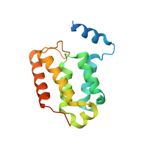
    Infection by Leishmania and Trypanosoma causes severe disease and can be fatal. The reduced effectiveness of current treatments is largely due to drug resistance, hence the urgent need to develop new drugs, preferably against novel targets. We have recently identified a mitochondrial membrane-anchored protein, designated MIX, which occurs exclusively in these parasites and is essential for virulence. We have determined the crystal structure of Leishmania major MIX to a resolution of 2.4 Å. MIX forms an all α-helical fold comprising seven α-helices that fold into a single domain. The distribution of helices is similar to a number of scaffold proteins, namely HEAT repeats, 14-3-3, and tetratricopeptide repeat proteins, suggesting that MIX mediates protein-protein interactions. Accordingly, using copurification and mass spectroscopy we were able to identify several proteins that may interact with MIX in vivo. Being parasite specific, MIX is a promising new drug target and, thus, the structure and potential interacting partners provide a basis for structure-guided drug discovery.


  • Organizational Affiliation
    • Biota Structural Biology Laboratory, St. Vincent's Institute of Medical Research, Victoria 3065, Australia.

Macromolecule Content 

  • Total Structure Weight: 141.45 kDa 
  • Atom Count: 7,963 
  • Modeled Residue Count: 936 
  • Deposited Residue Count: 1,232 
  • Unique protein chains: 1

Macromolecules

Find similar proteins by:|  3D Structure
Entity ID: 1
MoleculeChains  Sequence LengthOrganismDetailsImage
MIX1
A, B, C, D, E
A, B, C, D, E, F, G, H
154Leishmania majorMutation(s): 0 
Gene Names: LmjF08.1200
UniProt
Find proteins for Q4QI49 (Leishmania major)
Explore Q4QI49 
Go to UniProtKB:  Q4QI49
Entity Groups
Sequence Clusters30% Identity50% Identity70% Identity90% Identity95% Identity100% Identity
UniProt GroupQ4QI49
Sequence Annotations
Expand
Reference Sequence

Small Molecules

Ligands 1 Unique
IDChains Name / Formula / InChI Key2D Diagram3D Interactions
CO3

Query on CO3


Download:Ideal Coordinates CCD File
I [auth A]CARBONATE ION
C O3
BVKZGUZCCUSVTD-UHFFFAOYSA-L

Experimental Data & Validation

Experimental Data

  • Method: X-RAY DIFFRACTION
  • Resolution: 2.40 Å
  • R-Value Free:  0.261 (Depositor), 0.257 (DCC) 
  • R-Value Work:  0.212 (Depositor), 0.213 (DCC) 
  • R-Value Observed: 0.214 (Depositor) 
Space Group: P 21 21 21
Unit Cell:
Length ( Å )Angle ( ˚ )
a = 92.524α = 90
b = 100.389β = 90
c = 140.651γ = 90
Software Package:
Software NamePurpose
Blu-Icedata collection
SHARPphasing
PHENIXrefinement
HKL-2000data reduction
SCALAdata scaling

Structure Validation

View Full Validation Report


Entry History 

Deposition Data

Revision History  (Full details and data files)

  • Version 1.0: 2010-05-05
    Type: Initial release
  • Version 1.1: 2011-07-13
    Changes: Version format compliance
  • Version 1.2: 2024-02-21
    Changes: Data collection, Database references, Derived calculations