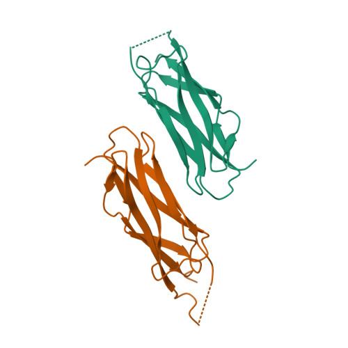
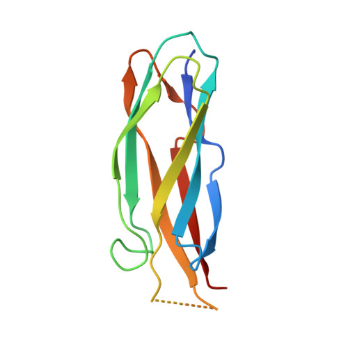
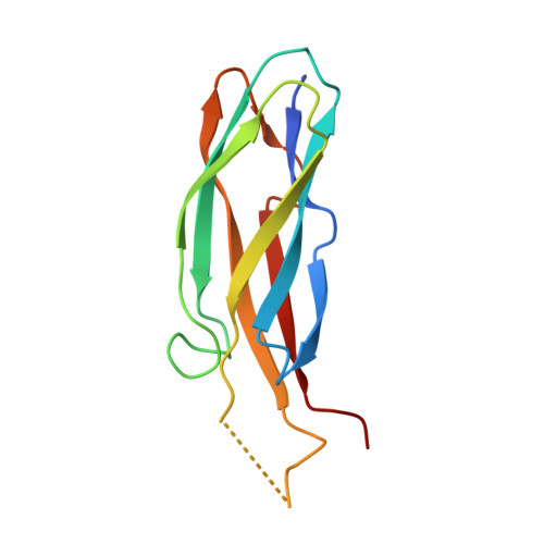
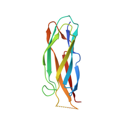
Structure of the Calx-beta domain of the integrin beta4 subunit: insights into function and cation-independent stability
Alonso-Garcia, N., Ingles-Prieto, A., Sonnenberg, A., de Pereda, J.M.(2009) Acta Crystallogr D Biol Crystallogr 65: 858-871
- PubMed: 19622870
- DOI: https://doi.org/10.1107/S0907444909018745
- Primary Citation of Related Structures:
3FQ4, 3FSO, 3H6A - PubMed Abstract:
The integrin alpha6beta4 is a receptor for laminins and provides stable adhesion of epithelial cells to the basement membranes. In addition, alpha6beta4 is important for keratinocyte migration during wound healing and favours the invasion of carcinomas into surrounding tissue. The cytoplasmic domain of the beta4 subunit is responsible for most of the intracellular interactions of the integrin; it contains four fibronectin type III domains and a Calx-beta motif. The crystal structure of the Calx-beta domain of beta4 was determined to 1.48 A resolution. The structure does not contain cations and biophysical data support the supposition that the Calx-beta domain of beta4 does not bind calcium. Comparison of the Calx-beta domain of beta4 with the calcium-binding domains of Na(+)/Ca(2+)-exchanger 1 reveals that in beta4 Arg1003 occupies a position equivalent to that of the calcium ions in the Na(+)/Ca(2+)-exchanger. By combining mutagenesis and thermally induced unfolding, it is shown that Arg1003 contributes to the stability of the Calx-beta domain. The structure of the Calx-beta domain is discussed in the context of the function and intracellular interactions of the integrin beta4 subunit and a putative functional site is proposed.
Organizational Affiliation:
Instituto de Biología Molecular y Celular del Cáncer, Consejo Superior de Investigaciones Científicas-Universidad de Salamanca, Campus Unamuno, 37007 Salamanca, Spain.


