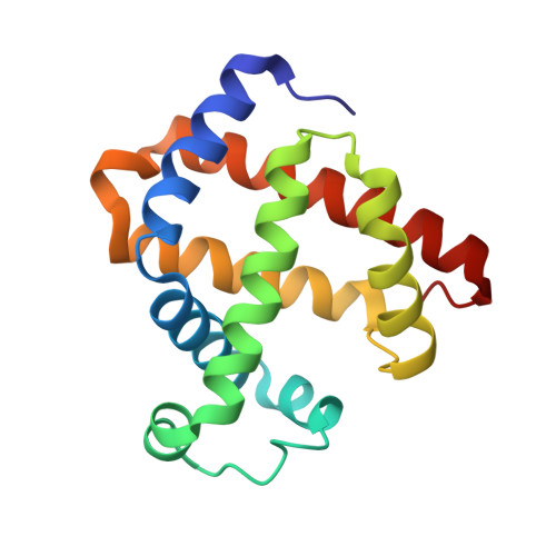
Optical detection of disordered water within a protein cavity.
Goldbeck, R.A., Pillsbury, M.L., Jensen, R.A., Mendoza, J.L., Nguyen, R.L., Olson, J.S., Soman, J., Kliger, D.S., Esquerra, R.M.(2009) J Am Chem Soc 131: 12265-12272
- PubMed: 19655795
- DOI: https://doi.org/10.1021/ja903409j
- Primary Citation of Related Structures:
3H57, 3H58 - PubMed Abstract:
Internal water molecules are important to protein structure and function, but positional disorder and low occupancies can obscure their detection by X-ray crystallography. Here, we show that water can be detected within the distal cavities of myoglobin mutants by subtle changes in the absorbance spectrum of pentacoordinate heme, even when the presence of solvent is not readily observed in the corresponding crystal structures. A well-defined, noncoordinated water molecule hydrogen bonded to the distal histidine (His64) is seen within the distal heme pocket in the crystal structure of wild type (wt) deoxymyoglobin. Displacement of this water decreases the rate of ligand entry into wt Mb, and we have shown previously that the entry of this water is readily detected optically after laser photolysis of MbCO complexes. However, for L29F and V68L Mb no discrete positions for solvent molecules are seen in the electron density maps of the crystal structures even though His64 is still present and slow rates of ligand binding indicative of internal water are observed. In contrast, time-resolved perturbations of the visible absorption bands of L29F and V68L deoxyMb generated after laser photolysis detect the entry and significant occupancy of water within the distal pockets of these variants. Thus, the spectral perturbation of pentacoordinate heme offers a potentially robust system for measuring nonspecific hydration of the active sites of heme proteins.
- Department of Chemistry and Biochemistry, University of California, Santa Cruz, California 95064, USA. goldbeck@chemistry.ucsc.edu
Organizational Affiliation:


