


Structure of YqeH GTPase from Bacillus anthracis with dGDP Bound
Brunzelle, J.S., Anderson, S.M., Wawrzak, Z., Xu, U., Cui, H., Savchenko, A., Anderson, W.F.To be published.
Experimental Data Snapshot
Starting Model: experimental
View more details
Entity ID: 1 | |||||
|---|---|---|---|---|---|
| Molecule | Chains | Sequence Length | Organism | Details | Image |
| GTPase family protein | 368 | Bacillus anthracis str. Sterne | Mutation(s): 0 Gene Names: BAS4233, BA_4562, GBAA4562, GBAA_4562, YqeH | 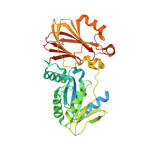 | |
UniProt | |||||
Entity Groups | |||||
| Sequence Clusters | 30% Identity50% Identity70% Identity90% Identity95% Identity100% Identity | ||||
| UniProt Group | A0A1J9WBB5 | ||||
Sequence AnnotationsExpand | |||||
Reference Sequence | |||||
| Ligands 1 Unique | |||||
|---|---|---|---|---|---|
| ID | Chains | Name / Formula / InChI Key | 2D Diagram | 3D Interactions | |
| DGI Download:Ideal Coordinates CCD File | B [auth A] | 2'-DEOXYGUANOSINE-5'-DIPHOSPHATE C10 H15 N5 O10 P2 CIKGWCTVFSRMJU-KVQBGUIXSA-N |  | ||
| Modified Residues 1 Unique | |||||
|---|---|---|---|---|---|
| ID | Chains | Type | Formula | 2D Diagram | Parent |
| MSE Query on MSE | A | L-PEPTIDE LINKING | C5 H11 N O2 Se |  | MET |
| Length ( Å ) | Angle ( ˚ ) |
|---|---|
| a = 83.13 | α = 90 |
| b = 58.99 | β = 99.38 |
| c = 77.72 | γ = 90 |
| Software Name | Purpose |
|---|---|
| BLU-MAX | data collection |
| MOLREP | phasing |
| REFMAC | refinement |
| XDS | data reduction |
| XSCALE | data scaling |


