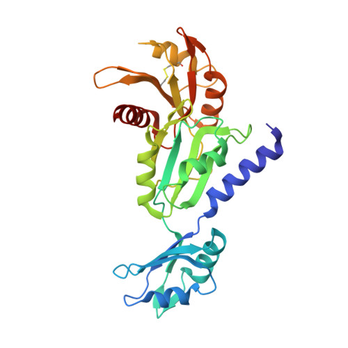
3H2U

Human raver1 RRM1, RRM2, and RRM3 domains in complex with human vinculin tail domain Vt


Experimental Data Snapshot

  • Method: X-RAY DIFFRACTION
  • Resolution: 2.75 Å
  • R-Value Free: 0.274 
  • R-Value Work: 0.225 
  • R-Value Observed: 0.227 

Starting Model: experimental
View more details

wwPDB Validation   3D Report Full Report


This is version 1.3 of the entry. See complete history


Literature

Raver1 interactions with vinculin and RNA suggest a feed-forward pathway in directing mRNA to focal adhesions

Lee, J.H.Rangarajan, E.S.Yogesha, S.D.Izard, T.

(2009) Structure 17: 833-842

  • DOI: https://doi.org/10.1016/j.str.2009.04.010
  • Primary Citation of Related Structures:  
    3H2U, 3H2V

  • PubMed Abstract: 

    The translational machinery of the cell relocalizes to focal adhesions following the activation of integrin receptors. This response allows for rapid, local production of components needed for adhesion complex assembly and signaling. Vinculin links focal adhesions to the actin cytoskeleton following its activation by integrin signaling, which severs intramolecular interactions of vinculin's head and tail (Vt) domains. Our vinculin:raver1 crystal structures and binding studies show that activated Vt selectively interacts with one of the three RNA recognition motifs of raver1, that the vinculin:raver1 complex binds to F-actin, and that raver1 binds selectively to RNA, including a sequence found in vinculin mRNA. Further, mutation of residues that mediate interaction of raver1 with vinculin abolish their colocalization in cells. These findings suggest a feed-forward model where vinculin activation at focal adhesions provides a scaffold for recruitment of raver1 and its mRNA cargo to facilitate the production of components of adhesion complexes.


  • Organizational Affiliation

    Cell Adhesion Laboratory, Department of Cancer Biology, The Scripps Research Institute, Jupiter, FL 33458, USA.


Macromolecules
Find similar proteins by:  (by identity cutoff)  |  3D Structure
Entity ID: 1
MoleculeChains Sequence LengthOrganismDetailsImage
Vinculin
A, C
188Homo sapiensMutation(s): 0 
UniProt & NIH Common Fund Data Resources
Find proteins for P18206 (Homo sapiens)
Explore P18206 
Go to UniProtKB:  P18206
PHAROS:  P18206
GTEx:  ENSG00000035403 
Entity Groups  
Sequence Clusters30% Identity50% Identity70% Identity90% Identity95% Identity100% Identity
UniProt GroupP18206
Sequence Annotations
Expand
  • Reference Sequence
Find similar proteins by:  (by identity cutoff)  |  3D Structure
Entity ID: 2
MoleculeChains Sequence LengthOrganismDetailsImage
Raver-1
B, D
283Homo sapiensMutation(s): 0 
Gene Names: RAVER1KIAA1978
UniProt & NIH Common Fund Data Resources
Find proteins for Q8IY67 (Homo sapiens)
Explore Q8IY67 
Go to UniProtKB:  Q8IY67
PHAROS:  Q8IY67
Entity Groups  
Sequence Clusters30% Identity50% Identity70% Identity90% Identity95% Identity100% Identity
UniProt GroupQ8IY67
Sequence Annotations
Expand
  • Reference Sequence
Experimental Data & Validation

Experimental Data

  • Method: X-RAY DIFFRACTION
  • Resolution: 2.75 Å
  • R-Value Free: 0.274 
  • R-Value Work: 0.225 
  • R-Value Observed: 0.227 
  • Space Group: P 21 21 21
Unit Cell:
Length ( Å )Angle ( ˚ )
a = 86.754α = 90
b = 94.906β = 90
c = 186.027γ = 90
Software Package:
Software NamePurpose
BUSTER-TNTrefinement

Structure Validation

View Full Validation Report


Entry History 

Deposition Data

Revision History  (Full details and data files)

  • Version 1.0: 2009-07-28
    Type: Initial release
  • Version 1.1: 2011-07-13
    Changes: Version format compliance
  • Version 1.2: 2023-09-06
    Changes: Data collection, Database references, Refinement description
  • Version 1.3: 2024-10-16
    Changes: Structure summary