3GYR | pdb_00003gyr

Structure of Phenoxazinone synthase from Streptomyces antibioticus reveals a new type 2 copper center.


Experimental Data Snapshot

  • Method: X-RAY DIFFRACTION
  • Resolution: 2.30 Å
  • R-Value Free: 
    0.222 (Depositor), 0.222 (DCC) 
  • R-Value Work: 
    0.165 (Depositor), 0.165 (DCC) 
  • R-Value Observed: 
    0.167 (Depositor) 

Starting Model: experimental
View more details

wwPDB Validation 3D Report Full Report

Validation slider image for 3GYR

This is version 1.5 of the entry. See complete history

Literature

Structure of Phenoxazinone Synthase from Streptomyces Antibioticus Reveals a New Type 2 Copper Center.

Smith, A.W.Camara-Artigas, A.Wang, M.Allen, J.P.Francisco, W.A.

(2006) Biochemistry 45: 4378

  • DOI: https://doi.org/10.1021/bi0525526
  • Primary Citation Related Structures: 
    3GYR

  • PubMed Abstract: 

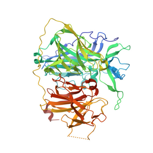
    The multicopper oxidase phenoxazinone synthase (PHS) catalyzes the penultimate step in the biosynthesis of the antibiotic actinomycin D by Streptomyces antibioticus. PHS exists in two oligomeric forms: a dimeric form and a hexameric form, with older actinomycin-producing cultures containing predominately the hexameric form. The structure of hexameric PHS has been determined using X-ray diffraction to a resolution limit of 2.30 A and is found to contain several unexpected and distinctive features. The structure forms a hexameric ring that is centered on a pseudo 6-fold axis and has an outer diameter of 185 A with a large central cavity that has a diameter of 50 A. This hexameric structure is stabilized by a long loop connecting two domains; bound to this long loop is a fifth copper atom that is present as a type 2 copper. This copper atom is not present in any other multicopper oxidase, and its presence appears to stabilize the hexameric structure.


  • Organizational Affiliation
    • Department of Chemistry and Biochemistry, Arizona State University, Tempe Arizona 85287-1604, USA.

Macromolecule Content 

  • Total Structure Weight: 810.24 kDa 
  • Atom Count: 59,241 
  • Modeled Residue Count: 7,044 
  • Deposited Residue Count: 7,344 
  • Unique protein chains: 1

Macromolecules

Find similar proteins by:|  3D Structure
Entity ID: 1
MoleculeChains  Sequence LengthOrganismDetailsImage
Phenoxazinone synthase
A, B, C, D, E
A, B, C, D, E, F, G, H, I, J, K, L
612Streptomyces antibioticusMutation(s): 1 
Gene Names: phsA
EC: 1 (PDB Primary Data), 1.10.3.4 (UniProt)
UniProt
Find proteins for Q53692 (Streptomyces antibioticus)
Explore Q53692 
Go to UniProtKB:  Q53692
Entity Groups
Sequence Clusters30% Identity50% Identity70% Identity90% Identity95% Identity100% Identity
UniProt GroupQ53692
Sequence Annotations
Expand
Reference Sequence

Small Molecules

Ligands 3 Unique
IDChains Name / Formula / InChI Key2D Diagram3D Interactions
C2O

Query on C2O


Download:Ideal Coordinates CCD File
DB [auth I]
EA [auth D]
IB [auth J]
JA [auth E]
NB [auth K]
DB [auth I],
EA [auth D],
IB [auth J],
JA [auth E],
NB [auth K],
OA [auth F],
P [auth A],
SB [auth L],
TA [auth G],
U [auth B],
YA [auth H],
Z [auth C]
CU-O-CU LINKAGE
Cu2 O
BERDEBHAJNAUOM-UHFFFAOYSA-N
GOL

Query on GOL


Download:Ideal Coordinates CCD File
AA [auth C]
EB [auth I]
FA [auth D]
JB [auth J]
KA [auth E]
AA [auth C],
EB [auth I],
FA [auth D],
JB [auth J],
KA [auth E],
OB [auth K],
PA [auth F],
Q [auth A],
TB [auth L],
UA [auth G],
V [auth B],
ZA [auth H]
GLYCEROL
C3 H8 O3
PEDCQBHIVMGVHV-UHFFFAOYSA-N
CU

Query on CU


Download:Ideal Coordinates CCD File
AB [auth I]
BA [auth D]
BB [auth I]
CA [auth D]
CB [auth I]
AB [auth I],
BA [auth D],
BB [auth I],
CA [auth D],
CB [auth I],
DA [auth D],
FB [auth J],
GA [auth E],
GB [auth J],
HA [auth E],
HB [auth J],
IA [auth E],
KB [auth K],
LA [auth F],
LB [auth K],
M [auth A],
MA [auth F],
MB [auth K],
N [auth A],
NA [auth F],
O [auth A],
PB [auth L],
QA [auth G],
QB [auth L],
R [auth B],
RA [auth G],
RB [auth L],
S [auth B],
SA [auth G],
T [auth B],
VA [auth H],
W [auth C],
WA [auth H],
X [auth C],
XA [auth H],
Y [auth C]
COPPER (II) ION
Cu
JPVYNHNXODAKFH-UHFFFAOYSA-N

Experimental Data & Validation

Experimental Data

  • Method: X-RAY DIFFRACTION
  • Resolution: 2.30 Å
  • R-Value Free:  0.222 (Depositor), 0.222 (DCC) 
  • R-Value Work:  0.165 (Depositor), 0.165 (DCC) 
  • R-Value Observed: 0.167 (Depositor) 
Space Group: P 1
Unit Cell:
Length ( Å )Angle ( ˚ )
a = 109.494α = 117.04
b = 163.456β = 95.74
c = 164.352γ = 107.23
Software Package:
Software NamePurpose
REFMACrefinement
CNSrefinement
CrystalCleardata collection
MOSFLMdata reduction
CCP4data scaling
CNSphasing

Structure Validation

View Full Validation Report


Entry History 

Deposition Data

Revision History  (Full details and data files)

  • Version 1.0: 2009-04-28
    Type: Initial release
  • Version 1.1: 2011-07-13
    Changes: Advisory, Derived calculations, Refinement description, Version format compliance
  • Version 1.2: 2018-04-04
    Changes: Data collection
  • Version 1.3: 2019-07-24
    Changes: Data collection, Refinement description
  • Version 1.4: 2021-10-13
    Changes: Database references, Derived calculations
  • Version 1.5: 2023-09-06
    Changes: Data collection, Refinement description