3GTD | pdb_00003gtd

2.4 Angstrom crystal structure of fumarate hydratase from Rickettsia prowazekii


Experimental Data Snapshot

  • Method: X-RAY DIFFRACTION
  • Resolution: 2.40 Å
  • R-Value Free: 
    0.226 (Depositor), 0.229 (DCC) 
  • R-Value Work: 
    0.193 (Depositor), 0.199 (DCC) 
  • R-Value Observed: 
    0.194 (Depositor) 

Starting Model: experimental
View more details

wwPDB Validation 3D Report Full Report

Validation slider image for 3GTD

This is version 1.3 of the entry. See complete history

Literature

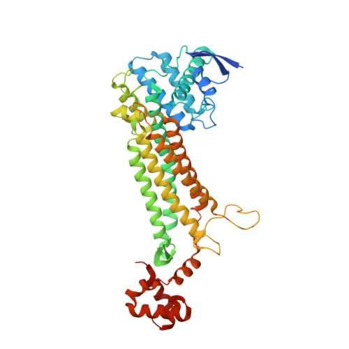
Structure of fumarate hydratase from Rickettsia prowazekii, the agent of typhus and suspected relative of the mitochondria.

Phan, I.Subramanian, S.Olsen, C.Edwards, T.E.Guo, W.Zhang, Y.Van Voorhis, W.C.Stewart, L.J.Myler, P.J.

(2011) Acta Crystallogr Sect F Struct Biol Cryst Commun 67: 1123-1128

  • DOI: https://doi.org/10.1107/S174430911102690X
  • Primary Citation Related Structures: 
    3GTD

  • PubMed Abstract: 

    Rickettsiae are obligate intracellular parasites of eukaryotic cells that are the causative agents responsible for spotted fever and typhus. Their small genome (about 800 protein-coding genes) is highly conserved across species and has been postulated as the ancestor of the mitochondria. No genes that are required for glycolysis are found in the Rickettsia prowazekii or mitochondrial genomes, but a complete set of genes encoding components of the tricarboxylic acid cycle and the respiratory-chain complex is found in both. A 2.4 Å resolution crystal structure of R. prowazekii fumarate hydratase, an enzyme catalyzing the third step of the tricarboxylic acid cycle pathway that ultimately converts phosphoenolpyruvate into succinyl-CoA, has been solved. A structure alignment with human mitochondrial fumarate hydratase highlights the close similarity between R. prowazekii and mitochondrial enzymes.


  • Organizational Affiliation
    • Seattle Structural Genomics Center for Infectious Diseases (SSGCID), USA. isabelle.phan@sbri.org

Macromolecule Content 

  • Total Structure Weight: 106.94 kDa 
  • Atom Count: 6,784 
  • Modeled Residue Count: 855 
  • Deposited Residue Count: 964 
  • Unique protein chains: 1

Macromolecules

Find similar proteins by:|  3D Structure
Entity ID: 1
MoleculeChains  Sequence LengthOrganismDetailsImage
Fumarate hydratase class II
A, B
482Rickettsia prowazekii str. Madrid EMutation(s): 0 
Gene Names: fumCRP665
EC: 4.2.1.2
UniProt
Find proteins for Q9ZCQ4 (Rickettsia prowazekii (strain Madrid E))
Explore Q9ZCQ4 
Go to UniProtKB:  Q9ZCQ4
Entity Groups
Sequence Clusters30% Identity50% Identity70% Identity90% Identity95% Identity100% Identity
UniProt GroupQ9ZCQ4
Sequence Annotations
Expand
Reference Sequence

Experimental Data & Validation

Experimental Data

  • Method: X-RAY DIFFRACTION
  • Resolution: 2.40 Å
  • R-Value Free:  0.226 (Depositor), 0.229 (DCC) 
  • R-Value Work:  0.193 (Depositor), 0.199 (DCC) 
  • R-Value Observed: 0.194 (Depositor) 
Space Group: P 31 2 1
Unit Cell:
Length ( Å )Angle ( ˚ )
a = 144.9α = 90
b = 144.9β = 90
c = 106.21γ = 120
Software Package:
Software NamePurpose
ADSCdata collection
PHASERphasing
REFMACrefinement
XDSdata reduction
XSCALEdata scaling

Structure Validation

View Full Validation Report


Entry History 

Deposition Data

Revision History  (Full details and data files)

  • Version 1.0: 2009-07-14
    Type: Initial release
  • Version 1.1: 2011-07-13
    Changes: Advisory, Version format compliance
  • Version 1.2: 2011-09-21
    Changes: Database references
  • Version 1.3: 2023-09-06
    Changes: Data collection, Database references, Derived calculations, Refinement description