3GPE | pdb_00003gpe

Crystal Structure Analysis of PKC (alpha)-C2 domain complexed with Ca2+ and PtdIns(4,5)P2


Experimental Data Snapshot

  • Method: X-RAY DIFFRACTION
  • Resolution: 2.00 Å
  • R-Value Free: 
    0.277 (Depositor), 0.284 (DCC) 
  • R-Value Work: 
    0.243 (Depositor), 0.243 (DCC) 
  • R-Value Observed: 
    0.244 (Depositor) 

Starting Model: experimental
View more details

wwPDB Validation 3D Report Full Report

Validation slider image for 3GPE

Ligand Structure Quality Assessment 


This is version 1.3 of the entry. See complete history

Literature

Structural and mechanistic insights into the association of PKCalpha-C2 domain to PtdIns(4,5)P2.

Guerrero-Valero, M.Ferrer-Orta, C.Querol-Audi, J.Marin-Vicente, C.Fita, I.Gomez-Fernandez, J.C.Verdaguer, N.Corbalan-Garcia, S.

(2009) Proc Natl Acad Sci U S A 106: 6603-6607

  • DOI: https://doi.org/10.1073/pnas.0813099106
  • Primary Citation Related Structures: 
    3GPE

  • PubMed Abstract: 

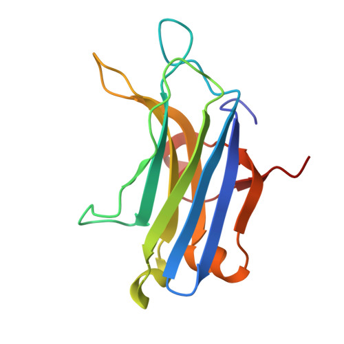
    C2 domains are widely-spread protein signaling motifs that in classical PKCs act as Ca(2+)-binding modules. However, the molecular mechanisms of their targeting process at the plasma membrane remain poorly understood. Here, the crystal structure of PKCalpha-C2 domain in complex with Ca(2+), 1,2-dihexanoyl-sn-glycero-3-[phospho-L-serine] (PtdSer), and 1,2-diayl-sn-glycero-3-[phosphoinositol-4,5-bisphosphate] [PtdIns(4,5)P(2)] shows that PtdSer binds specifically to the calcium-binding region, whereas PtdIns(4,5)P(2) occupies the concave surface of strands beta3 and beta4. Strikingly, the structure reveals a PtdIns(4,5)P(2)-C2 domain-binding mode in which the aromatic residues Tyr-195 and Trp-245 establish direct interactions with the phosphate moieties of the inositol ring. Mutations that abrogate Tyr-195 and Trp-245 recognition of PtdIns(4,5)P(2) severely impaired the ability of PKCalpha to localize to the plasma membrane. Notably, these residues are highly conserved among C2 domains of topology I, and a general mechanism of C2 domain-membrane docking mediated by PtdIns(4,5)P(2) is presented.


  • Organizational Affiliation
    • Department of Bioquímica y Biología Molecular A, Facultad de Veterinaria, Universidad de Murcia, 30100 Murcia, Spain.

Macromolecule Content 

  • Total Structure Weight: 17.31 kDa 
  • Atom Count: 1,248 
  • Modeled Residue Count: 137 
  • Deposited Residue Count: 137 
  • Unique protein chains: 1

Macromolecules

Find similar proteins by:|  3D Structure
Entity ID: 1
MoleculeChains  Sequence LengthOrganismDetailsImage
Protein kinase C alpha type137Rattus norvegicusMutation(s): 0 
Gene Names: PrkcaPkca
EC: 2.7.11.13
UniProt
Find proteins for P05696 (Rattus norvegicus)
Explore P05696 
Go to UniProtKB:  P05696
Entity Groups
Sequence Clusters30% Identity50% Identity70% Identity90% Identity95% Identity100% Identity
UniProt GroupP05696
Sequence Annotations
Expand
Reference Sequence

Experimental Data & Validation

Experimental Data

  • Method: X-RAY DIFFRACTION
  • Resolution: 2.00 Å
  • R-Value Free:  0.277 (Depositor), 0.284 (DCC) 
  • R-Value Work:  0.243 (Depositor), 0.243 (DCC) 
  • R-Value Observed: 0.244 (Depositor) 
Space Group: P 32 2 1
Unit Cell:
Length ( Å )Angle ( ˚ )
a = 57.848α = 90
b = 57.848β = 90
c = 90.479γ = 120
Software Package:
Software NamePurpose
ADSCdata collection
AMoREphasing
REFMACrefinement
MOSFLMdata reduction
SCALAdata scaling

Structure Validation

View Full Validation Report


Ligand Structure Quality Assessment 


Entry History 

Deposition Data

Revision History  (Full details and data files)

  • Version 1.0: 2009-05-05
    Type: Initial release
  • Version 1.1: 2011-07-13
    Changes: Advisory, Version format compliance
  • Version 1.2: 2024-02-21
    Changes: Advisory, Data collection, Database references, Derived calculations, Structure summary
  • Version 1.3: 2024-04-03
    Changes: Refinement description