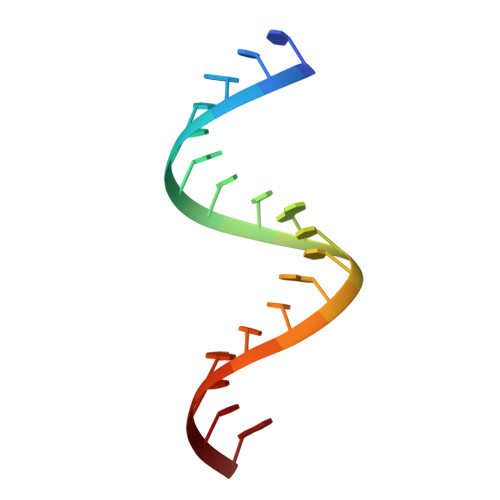
Structural insights into CUG repeats containing the 'stretched U-U wobble': implications for myotonic dystrophy.
Kiliszek, A., Kierzek, R., Krzyzosiak, W.J., Rypniewski, W.(2009) Nucleic Acids Res 37: 4149-4156
- PubMed: 19433512
- DOI: https://doi.org/10.1093/nar/gkp350
- Primary Citation of Related Structures:
3GLP, 3GM7 - PubMed Abstract:
Tracks containing CUG repeats are abundant in human gene transcripts. Their biological role includes modulation of pre-mRNA splicing, mRNA transport and regulation of translation. Expanded forms of CUG runs are associated with pathogenesis of several neurodegenerative diseases, including myotonic dystrophy type 1. We have analysed two crystal structures of RNA duplexes containing the CUG repeats: G(CUG)(2)C and (CUG)(6). The first of the structures, analysed at 1.23 A resolution, is of an oligomer designed by us. The second model was obtained after 'detwinning' the 1.58 A X-ray data previously deposited in the PDB. The RNA duplexes are in the A-form in which all the C-G pairs form Watson-Crick interactions while all the uridine pairs can be described as U*U cis wobble having only one hydrogen bond between the bases. The residue, which accepts the H-bond, is inclined towards the minor groove. This previously unreported base pairing can be described as 'stretched U-U wobble'. The regular hydrogen-bonding pattern of interactions with the solvent, the electrostatic charge distribution and surface features indicate the ligand binding potential of the CUG tracks.
- Institute of Bioorganic Chemistry, Polish Academy of Sciences, Noskowskiego 12/14, 61-704 Poznan, Poland.
Organizational Affiliation:


