3GLH | pdb_00003glh

Crystal Structure of the E. coli clamp loader bound to Psi Peptide


Experimental Data Snapshot

  • Method: X-RAY DIFFRACTION
  • Resolution: 3.89 Å
  • R-Value Free: 
    0.361 (Depositor), 0.348 (DCC) 
  • R-Value Work: 
    0.359 (Depositor), 0.347 (DCC) 

wwPDB Validation 3D Report Full Report

Validation slider image for 3GLH

This is version 1.3 of the entry. See complete history

Literature

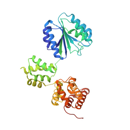
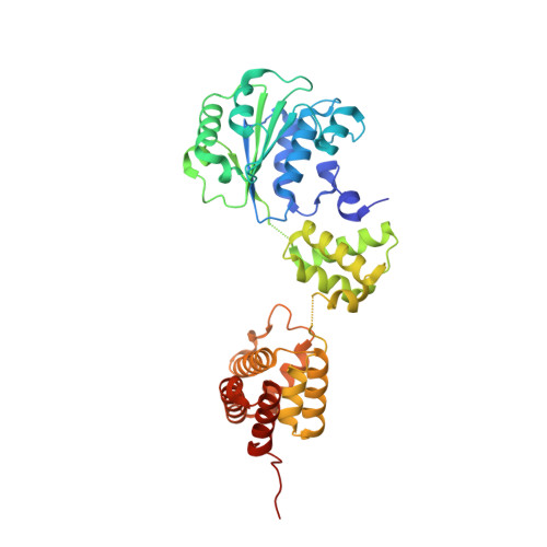
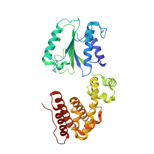
The mechanism of ATP-dependent primer-template recognition by a clamp loader complex.

Simonetta, K.R.Kazmirski, S.L.Goedken, E.R.Cantor, A.J.Kelch, B.A.McNally, R.Seyedin, S.N.Makino, D.L.O'Donnell, M.Kuriyan, J.

(2009) Cell 137: 659-671

  • DOI: https://doi.org/10.1016/j.cell.2009.03.044
  • Primary Citation Related Structures: 
    3GLF, 3GLG, 3GLH, 3GLI

  • PubMed Abstract: 

    Clamp loaders load sliding clamps onto primer-template DNA. The structure of the E. coli clamp loader bound to DNA reveals the formation of an ATP-dependent spiral of ATPase domains that tracks only the template strand, allowing recognition of both RNA and DNA primers. Unlike hexameric helicases, in which DNA translocation requires distinct conformations of the ATPase domains, the clamp loader spiral is symmetric and is set up to trigger release upon DNA recognition. Specificity for primed DNA arises from blockage of the end of the primer and accommodation of the emerging template along a surface groove. A related structure reveals how the psi protein, essential for coupling the clamp loader to single-stranded DNA-binding protein (SSB), binds to the clamp loader. By stabilizing a conformation of the clamp loader that is consistent with the ATPase spiral observed upon DNA binding, psi binding promotes the clamp-loading activity of the complex.


  • Organizational Affiliation
    • Department of Molecular and Cell Biology, Howard Hughes Medical Institute, University of California, Berkeley, Berkeley, CA 94720, USA.

Macromolecule Content 

  • Total Structure Weight: 603.41 kDa 
  • Atom Count: 41,322 
  • Modeled Residue Count: 5,277 
  • Deposited Residue Count: 5,415 
  • Unique protein chains: 3

Macromolecules

Find similar proteins by:|  3D Structure
Entity ID: 1
MoleculeChains  Sequence LengthOrganismDetailsImage
DNA polymerase III subunit delta
A, F, K
343Escherichia coli K-12Mutation(s): 0 
Gene Names: holAb0640JW0635
EC: 2.7.7.7
UniProt
Find proteins for P28630 (Escherichia coli (strain K12))
Explore P28630 
Go to UniProtKB:  P28630
Entity Groups
Sequence Clusters30% Identity50% Identity70% Identity90% Identity95% Identity100% Identity
UniProt GroupP28630
Sequence Annotations
Expand
Reference Sequence
Find similar proteins by:|  3D Structure
Entity ID: 2
MoleculeChains  Sequence LengthOrganismDetailsImage
DNA polymerase III subunit tau
B, C, D, G, H
B, C, D, G, H, I, L, M, N
376Escherichia coli K-12Mutation(s): 0 
Gene Names: dnaXdnaZdnaZXb0470JW0459
EC: 2.7.7.7
UniProt
Find proteins for P06710 (Escherichia coli (strain K12))
Explore P06710 
Go to UniProtKB:  P06710
Entity Groups
Sequence Clusters30% Identity50% Identity70% Identity90% Identity95% Identity100% Identity
UniProt GroupP06710
Sequence Annotations
Expand
Reference Sequence
Find similar proteins by:|  3D Structure
Entity ID: 3
MoleculeChains  Sequence LengthOrganismDetailsImage
DNA polymerase III subunit delta'
E, J, O
334Escherichia coli K-12Mutation(s): 0 
Gene Names: holBb1099JW1085
EC: 2.7.7.7
UniProt
Find proteins for P28631 (Escherichia coli (strain K12))
Explore P28631 
Go to UniProtKB:  P28631
Entity Groups
Sequence Clusters30% Identity50% Identity70% Identity90% Identity95% Identity100% Identity
UniProt GroupP28631
Sequence Annotations
Expand
Reference Sequence

Experimental Data & Validation

Experimental Data

  • Method: X-RAY DIFFRACTION
  • Resolution: 3.89 Å
  • R-Value Free:  0.361 (Depositor), 0.348 (DCC) 
  • R-Value Work:  0.359 (Depositor), 0.347 (DCC) 
Space Group: P 1 21 1
Unit Cell:
Length ( Å )Angle ( ˚ )
a = 100.162α = 90
b = 228.494β = 90
c = 164.74γ = 90
Software Package:
Software NamePurpose
HKL-2000data collection
AMoREphasing
PHENIXrefinement
HKL-2000data reduction
HKL-2000data scaling

Structure Validation

View Full Validation Report


Entry History 

Deposition Data

Revision History  (Full details and data files)

  • Version 1.0: 2009-05-26
    Type: Initial release
  • Version 1.1: 2011-07-13
    Changes: Version format compliance
  • Version 1.2: 2013-01-23
    Changes: Database references
  • Version 1.3: 2024-02-21
    Changes: Data collection, Database references