3GIL | pdb_00003gil

Dpo4 extension ternary complex with oxoG(anti)-T(anti) pair


Experimental Data Snapshot

  • Method: X-RAY DIFFRACTION
  • Resolution: 2.71 Å
  • R-Value Free: 
    0.259 (Depositor), 0.292 (DCC) 
  • R-Value Work: 
    0.200 (Depositor) 
  • R-Value Observed: 
    0.203 (Depositor) 

Starting Model: experimental
View more details

wwPDB Validation 3D Report Full Report

Validation slider image for 3GIL

Ligand Structure Quality Assessment 


This is version 1.4 of the entry. See complete history

Literature

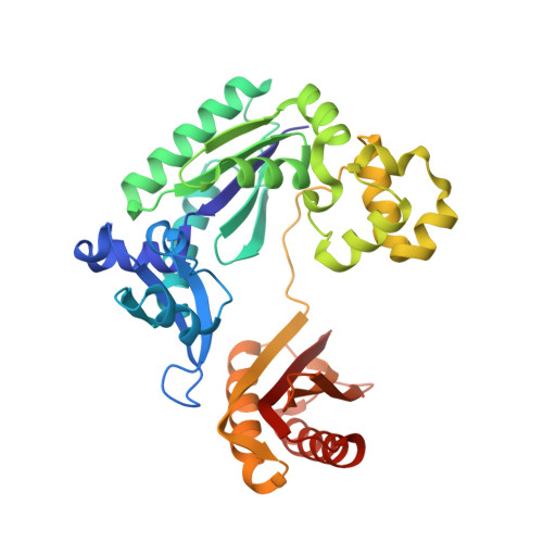
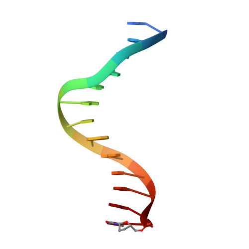
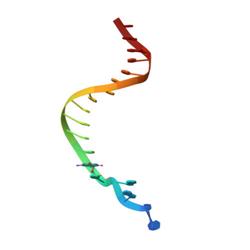
Impact of conformational heterogeneity of OxoG lesions and their pairing partners on bypass fidelity by Y family polymerases.

Rechkoblit, O.Malinina, L.Cheng, Y.Geacintov, N.E.Broyde, S.Patel, D.J.

(2009) Structure 17: 725-736

  • DOI: https://doi.org/10.1016/j.str.2009.03.011
  • Primary Citation Related Structures: 
    3GII, 3GIJ, 3GIK, 3GIL, 3GIM

  • PubMed Abstract: 

    7,8-Dihydro-8-oxoguanine (oxoG), the predominant oxidative DNA damage lesion, is processed differently by high-fidelity and Y-family lesion bypass polymerases. Although high-fidelity polymerases extend predominantly from an A base opposite an oxoG, the Y-family polymerases Dpo4 and human Pol eta preferentially extend from the oxoG*C base pair. We have determined crystal structures of extension Dpo4 ternary complexes with oxoG opposite C, A, G, or T and the next nascent base pair. We demonstrate that neither template backbone nor the architecture of the active site is perturbed by the oxoG(anti)*C and oxoG*A pairs. However, the latter manifest conformational heterogeneity, adopting both oxoG(syn)*A(anti) and oxoG(anti)*A(syn) alignment. Hence, the observed reduced primer extension from the dynamically flexible 3'-terminal primer base A is explained. Because of homology between Dpo4 and Pol eta, such a dynamic screening mechanism might be utilized by Dpo4 and Pol eta to regulate error-free versus error-prone bypass of oxoG and other lesions.


  • Organizational Affiliation
    • Structural Biology Program, Memorial Sloan-Kettering Cancer Center, New York, NY 10021, USA.

Macromolecule Content 

  • Total Structure Weight: 98.08 kDa 
  • Atom Count: 6,894 
  • Modeled Residue Count: 742 
  • Deposited Residue Count: 744 
  • Unique protein chains: 1
  • Unique nucleic acid chains: 2

Macromolecules


Find similar proteins by:|  3D Structure
Entity ID: 1
MoleculeChains  Sequence LengthOrganismDetailsImage
DNA polymerase IV
A, B
341Saccharolobus solfataricus P2Mutation(s): 0 
Gene Names: dbhdpo4SSO2448
EC: 2.7.7.7
UniProt
Find proteins for Q97W02 (Saccharolobus solfataricus (strain ATCC 35092 / DSM 1617 / JCM 11322 / P2))
Explore Q97W02 
Go to UniProtKB:  Q97W02
Entity Groups
Sequence Clusters30% Identity50% Identity70% Identity90% Identity95% Identity100% Identity
UniProt GroupQ97W02
Sequence Annotations
Expand
Reference Sequence
Find similar nucleic acids by:  Sequence
Entity ID: 2
MoleculeChains LengthOrganismImage
5'-D(*GP*TP*TP*GP*GP*AP*TP*GP*GP*TP*AP*GP*(2DT))-3'C [auth D],
E [auth H]
13N/A
Sequence Annotations
Expand
Reference Sequence
Find similar nucleic acids by:  Sequence
Entity ID: 3
MoleculeChains LengthOrganismImage
5'-D(*CP*TP*AP*AP*CP*(8OG)P*CP*TP*AP*CP*CP*AP*TP*CP*CP*AP*AP*C)-3'D [auth E],
F [auth J]
18N/A
Sequence Annotations
Expand
Reference Sequence

Small Molecules

Ligands 2 Unique
IDChains Name / Formula / InChI Key2D Diagram3D Interactions
DGT

Query on DGT


Download:Ideal Coordinates CCD File
G [auth A],
K [auth B]
2'-DEOXYGUANOSINE-5'-TRIPHOSPHATE
C10 H16 N5 O13 P3
HAAZLUGHYHWQIW-KVQBGUIXSA-N
CA

Query on CA


Download:Ideal Coordinates CCD File
H [auth A]
I [auth A]
J [auth A]
L [auth B]
M [auth B]
H [auth A],
I [auth A],
J [auth A],
L [auth B],
M [auth B],
N [auth B]
CALCIUM ION
Ca
BHPQYMZQTOCNFJ-UHFFFAOYSA-N

Experimental Data & Validation

Experimental Data

  • Method: X-RAY DIFFRACTION
  • Resolution: 2.71 Å
  • R-Value Free:  0.259 (Depositor), 0.292 (DCC) 
  • R-Value Work:  0.200 (Depositor) 
  • R-Value Observed: 0.203 (Depositor) 
Space Group: P 1 21 1
Unit Cell:
Length ( Å )Angle ( ˚ )
a = 53.289α = 90
b = 110.682β = 99.22
c = 102.19γ = 90
Software Package:
Software NamePurpose
DENZOdata reduction
SCALEPACKdata scaling
REFMACrefinement
PDB_EXTRACTdata extraction
HKL-2000data collection
HKL-2000data reduction
AMoREphasing

Structure Validation

View Full Validation Report


Ligand Structure Quality Assessment 


Entry History 

Deposition Data

Revision History  (Full details and data files)

  • Version 1.0: 2009-05-19
    Type: Initial release
  • Version 1.1: 2011-07-13
    Changes: Advisory, Version format compliance
  • Version 1.2: 2011-12-07
    Changes: Database references
  • Version 1.3: 2017-11-01
    Changes: Refinement description
  • Version 1.4: 2023-09-06
    Changes: Data collection, Database references, Derived calculations, Refinement description