


Crystal Structure of Helicobacter pylori Heme Oxygenase Hugz in Complex with Heme
Jiang, F., Hu, Y.L., Guo, Y., Guo, G., Zou, Q.M., Wang, D.C.To be published.
Experimental Data Snapshot
Entity ID: 1 | |||||
|---|---|---|---|---|---|
| Molecule | Chains | Sequence Length | Organism | Details | Image |
| heme oxygenase | 259 | Helicobacter pylori NCTC 11639 | Mutation(s): 0 Gene Names: HugZ EC: 1.14.99.3 (PDB Primary Data), 1.14.99 (UniProt) | 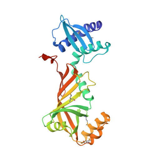 | |
UniProt | |||||
Entity Groups | |||||
| Sequence Clusters | 30% Identity50% Identity70% Identity90% Identity95% Identity100% Identity | ||||
| UniProt Group | O25087 | ||||
Sequence AnnotationsExpand | |||||
Reference Sequence | |||||
| Ligands 3 Unique | |||||
|---|---|---|---|---|---|
| ID | Chains | Name / Formula / InChI Key | 2D Diagram | 3D Interactions | |
| HEM Download:Ideal Coordinates CCD File | CA [auth E] G [auth A] GA [auth F] L [auth B] S [auth C] | PROTOPORPHYRIN IX CONTAINING FE C34 H32 Fe N4 O4 KABFMIBPWCXCRK-RGGAHWMASA-L |  | ||
| EDO Download:Ideal Coordinates CCD File | AA [auth D] BA [auth D] EA [auth E] FA [auth E] I [auth A] | 1,2-ETHANEDIOL C2 H6 O2 LYCAIKOWRPUZTN-UHFFFAOYSA-N |  | ||
| AZI Download:Ideal Coordinates CCD File | DA [auth E] H [auth A] HA [auth F] M [auth B] T [auth C] | AZIDE ION N3 IVRMZWNICZWHMI-UHFFFAOYSA-N |  | ||
| Modified Residues 1 Unique | |||||
|---|---|---|---|---|---|
| ID | Chains | Type | Formula | 2D Diagram | Parent |
| MSE Query on MSE | A, B, C, D, E A, B, C, D, E, F | L-PEPTIDE LINKING | C5 H11 N O2 Se |  | MET |
| Length ( Å ) | Angle ( ˚ ) |
|---|---|
| a = 88.33 | α = 90 |
| b = 139.34 | β = 90 |
| c = 152.9 | γ = 90 |
| Software Name | Purpose |
|---|---|
| SCALA | data scaling |
| SHARP | phasing |
| RESOLVE | phasing |
| CNS | refinement |
| PDB_EXTRACT | data extraction |
| ADSC | data collection |
| HKL-2000 | data reduction |


