3G9D | pdb_00003g9d

Crystal structure glycohydrolase


Experimental Data Snapshot

  • Method: X-RAY DIFFRACTION
  • Resolution: 2.50 Å
  • R-Value Free: 
    0.266 (Depositor), 0.263 (DCC) 
  • R-Value Work: 
    0.199 (Depositor) 
  • R-Value Observed: 
    0.202 (Depositor) 

Starting Model: experimental
View more details

wwPDB Validation 3D Report Full Report

Validation slider image for 3G9D

This is version 1.2 of the entry. See complete history

Literature

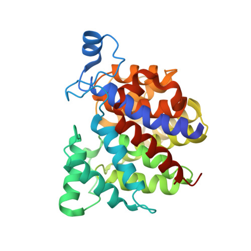
Crystal Structure of Dinitrogenase Reductase-activating Glycohydrolase (DRAG) Reveals Conservation in the ADP-Ribosylhydrolase Fold and Specific Features in the ADP-Ribose-binding Pocket

Li, X.-D.Huergo, L.F.Gasperina, A.Pedrosa, F.O.Merrick, M.Winkler, F.K.

(2009) J Mol Biology 390: 737-746

  • DOI: https://doi.org/10.1016/j.jmb.2009.05.031
  • Primary Citation Related Structures: 
    3G9D

  • PubMed Abstract: 

    Protein-reversible ADP-ribosylation is emerging as an important post-translational modification used to control enzymatic and protein activity in different biological systems. This modification regulates nitrogenase activity in several nitrogen-fixing bacterial species. ADP-ribosylation is catalyzed by ADP-ribosyltransferases and is reversed by ADP-ribosylhydrolases. The structure of the ADP-ribosylhydrolase that acts on Azospirillum brasilense nitrogenase (dinitrogenase reductase-activating glycohydrolase, DraG) has been solved at a resolution of 2.5 A. This bacterial member of the ADP-ribosylhydrolase family acts specifically towards a mono-ADP-ribosylated substrate. The protein shows an all-alpha-helix structure with two magnesium ions located in the active site. Comparison of the DraG structure with orthologues deposited in the Protein Data Bank from Archaea and mammals indicates that the ADP-ribosylhydrolase fold is conserved in all domains of life. Modeling of the binding of the substrate ADP-ribosyl moiety to DraG is in excellent agreement with biochemical data.


  • Organizational Affiliation
    • Biomolecular Research, Paul Scherrer Institut, Villigen, Switzerland.

Macromolecule Content 

  • Total Structure Weight: 64.98 kDa 
  • Atom Count: 4,518 
  • Modeled Residue Count: 580 
  • Deposited Residue Count: 594 
  • Unique protein chains: 1

Macromolecules

Find similar proteins by:|  3D Structure
Entity ID: 1
MoleculeChains  Sequence LengthOrganismDetailsImage
Dinitrogenase reductase activating glucohydrolase
A, B
297Azospirillum brasilenseMutation(s): 0 
Gene Names: draG
EC: 3.2.2.24
UniProt
Find proteins for A7XNI2 (Azospirillum brasilense)
Explore A7XNI2 
Go to UniProtKB:  A7XNI2
Entity Groups
Sequence Clusters30% Identity50% Identity70% Identity90% Identity95% Identity100% Identity
UniProt GroupA7XNI2
Sequence Annotations
Expand
Reference Sequence

Experimental Data & Validation

Experimental Data

  • Method: X-RAY DIFFRACTION
  • Resolution: 2.50 Å
  • R-Value Free:  0.266 (Depositor), 0.263 (DCC) 
  • R-Value Work:  0.199 (Depositor) 
  • R-Value Observed: 0.202 (Depositor) 
Space Group: P 65
Unit Cell:
Length ( Å )Angle ( ˚ )
a = 163.866α = 90
b = 163.866β = 90
c = 46.274γ = 120
Software Package:
Software NamePurpose
XDSdata scaling
PHASESphasing
REFMACrefinement
XDSdata reduction

Structure Validation

View Full Validation Report


Entry History 

Deposition Data

Revision History  (Full details and data files)

  • Version 1.0: 2009-07-14
    Type: Initial release
  • Version 1.1: 2011-07-13
    Changes: Version format compliance
  • Version 1.2: 2023-11-01
    Changes: Data collection, Database references, Derived calculations, Refinement description