3G7Z | pdb_00003g7z

CcdB dimer in complex with two C-terminal CcdA domains


Experimental Data Snapshot

  • Method: X-RAY DIFFRACTION
  • Resolution: 2.35 Å
  • R-Value Free: 
    0.272 (Depositor), 0.267 (DCC) 
  • R-Value Work: 
    0.203 (Depositor), 0.203 (DCC) 
  • R-Value Observed: 
    0.206 (Depositor) 

Starting Model: experimental
View more details

wwPDB Validation 3D Report Full Report

Validation slider image for 3G7Z

This is version 1.2 of the entry. See complete history

Literature

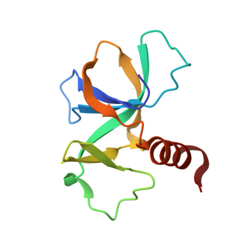
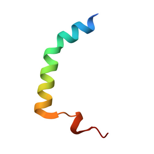
Rejuvenation of CcdB-Poisoned Gyrase by an Intrinsically Disordered Protein Domain.

De Jonge, N.Garcia-Pino, A.Buts, L.Haesaerts, S.Charlier, D.Zangger, K.Wyns, L.De Greve, H.Loris, R.

(2009) Mol Cell 35: 154-163

  • DOI: https://doi.org/10.1016/j.molcel.2009.05.025
  • Primary Citation Related Structures: 
    3G7Z, 3HPW

  • PubMed Abstract: 

    Toxin-antitoxin modules are small regulatory circuits that ensure survival of bacterial populations under challenging environmental conditions. The ccd toxin-antitoxin module on the F plasmid codes for the toxin CcdB and its antitoxin CcdA. CcdB poisons gyrase while CcdA actively dissociates CcdB:gyrase complexes in a process called rejuvenation. The CcdA:CcdB ratio modulates autorepression of the ccd operon. The mechanisms behind both rejuvenation and regulation of expression are poorly understood. We show that CcdA binds consecutively to two partially overlapping sites on CcdB, which differ in affinity by six orders of magnitude. The first, picomolar affinity interaction triggers a conformational change in CcdB that initiates the dissociation of CcdB:gyrase complexes by an allosteric segmental binding mechanism. The second, micromolar affinity binding event regulates expression of the ccd operon. Both functions of CcdA, rejuvenation and autoregulation, are mechanistically intertwined and depend crucially on the intrinsically disordered nature of the CcdA C-terminal domain.


  • Organizational Affiliation
    • Structural Biology Brussels, Vrije Universiteit Brussel, Pleinlaan 2, 1050 Brussels, Belgium.

Macromolecule Content 

  • Total Structure Weight: 32.2 kDa 
  • Atom Count: 2,205 
  • Modeled Residue Count: 269 
  • Deposited Residue Count: 274 
  • Unique protein chains: 2

Macromolecules

Find similar proteins by:|  3D Structure
Entity ID: 1
MoleculeChains  Sequence LengthOrganismDetailsImage
Cytotoxic protein ccdB
A, B
101Escherichia coliMutation(s): 0 
Gene Names: ccdBECOK12F043GletB
UniProt
Find proteins for P62554 (Escherichia coli (strain K12))
Explore P62554 
Go to UniProtKB:  P62554
Entity Groups
Sequence Clusters30% Identity50% Identity70% Identity90% Identity95% Identity100% Identity
UniProt GroupP62554
Sequence Annotations
Expand
Reference Sequence
Find similar proteins by:|  3D Structure
Entity ID: 2
MoleculeChains  Sequence LengthOrganismDetailsImage
Protein ccdA
C, D
36N/AMutation(s): 0 
UniProt
Find proteins for P62552 (Escherichia coli (strain K12))
Explore P62552 
Go to UniProtKB:  P62552
Entity Groups
Sequence Clusters30% Identity50% Identity70% Identity90% Identity95% Identity100% Identity
UniProt GroupP62552
Sequence Annotations
Expand
Reference Sequence

Experimental Data & Validation

Experimental Data

  • Method: X-RAY DIFFRACTION
  • Resolution: 2.35 Å
  • R-Value Free:  0.272 (Depositor), 0.267 (DCC) 
  • R-Value Work:  0.203 (Depositor), 0.203 (DCC) 
  • R-Value Observed: 0.206 (Depositor) 
Space Group: P 1 21 1
Unit Cell:
Length ( Å )Angle ( ˚ )
a = 58.248α = 90
b = 38.377β = 94.68
c = 62.498γ = 90
Software Package:
Software NamePurpose
ADSCdata collection
PHASERphasing
PHENIXrefinement
DENZOdata reduction
SCALEPACKdata scaling

Structure Validation

View Full Validation Report


Entry History 

Deposition Data

Revision History  (Full details and data files)

  • Version 1.0: 2009-08-11
    Type: Initial release
  • Version 1.1: 2011-07-13
    Changes: Version format compliance
  • Version 1.2: 2023-09-06
    Changes: Data collection, Database references, Refinement description