


Crystal structure of trans-aconitate 3-methyltransferase from yeast
Burgie, E.S., Bingman, C.A., Wesenberg, G.E., Phillips Jr., G.N.To be published.
Experimental Data Snapshot
Entity ID: 1 | |||||
|---|---|---|---|---|---|
| Molecule | Chains | Sequence Length | Organism | Details | Image |
| Trans-aconitate 3-methyltransferase | 299 | Saccharomyces cerevisiae | Mutation(s): 0 Gene Names: P32643, S000000977, SYGP-ORF63, TAM1, TMT1, YER175C EC: 2.1.1.145 | 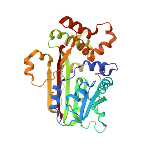 | |
UniProt | |||||
Entity Groups | |||||
| Sequence Clusters | 30% Identity50% Identity70% Identity90% Identity95% Identity100% Identity | ||||
| UniProt Group | P32643 | ||||
Sequence AnnotationsExpand | |||||
Reference Sequence | |||||
| Ligands 4 Unique | |||||
|---|---|---|---|---|---|
| ID | Chains | Name / Formula / InChI Key | 2D Diagram | 3D Interactions | |
| SAH Download:Ideal Coordinates CCD File | J [auth A] | S-ADENOSYL-L-HOMOCYSTEINE C14 H20 N6 O5 S ZJUKTBDSGOFHSH-WFMPWKQPSA-N |  | ||
| T8N Download:Ideal Coordinates CCD File | K [auth A] | (2E)-2-(2-methoxy-2-oxoethyl)but-2-enedioic acid C7 H8 O6 MRNZYUAGJLJQAM-DUXPYHPUSA-N |  | ||
| DMS Download:Ideal Coordinates CCD File | L [auth A] | DIMETHYL SULFOXIDE C2 H6 O S IAZDPXIOMUYVGZ-UHFFFAOYSA-N |  | ||
| EDO Download:Ideal Coordinates CCD File | B [auth A] C [auth A] D [auth A] E [auth A] F [auth A] | 1,2-ETHANEDIOL C2 H6 O2 LYCAIKOWRPUZTN-UHFFFAOYSA-N |  | ||
| Modified Residues 1 Unique | |||||
|---|---|---|---|---|---|
| ID | Chains | Type | Formula | 2D Diagram | Parent |
| MSE Query on MSE | A | L-PEPTIDE LINKING | C5 H11 N O2 Se |  | MET |
| Length ( Å ) | Angle ( ˚ ) |
|---|---|
| a = 36.834 | α = 90 |
| b = 91.891 | β = 90 |
| c = 104.269 | γ = 90 |
| Software Name | Purpose |
|---|---|
| DENZO | data reduction |
| SCALEPACK | data scaling |
| SHARP | phasing |
| DM | phasing |
| REFMAC | refinement |
| PDB_EXTRACT | data extraction |
| Blu-Ice | data collection |
| HKL-2000 | data reduction |
| HKL-2000 | data scaling |


