3G4T

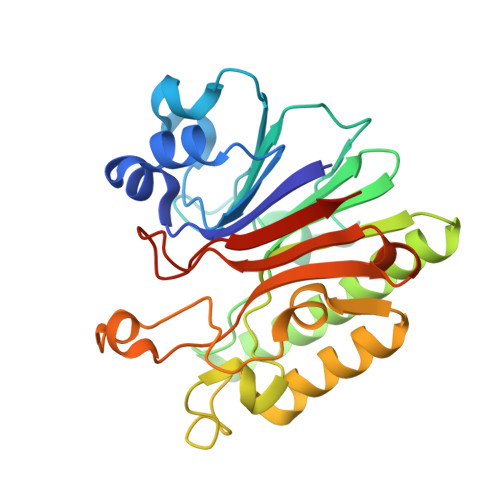
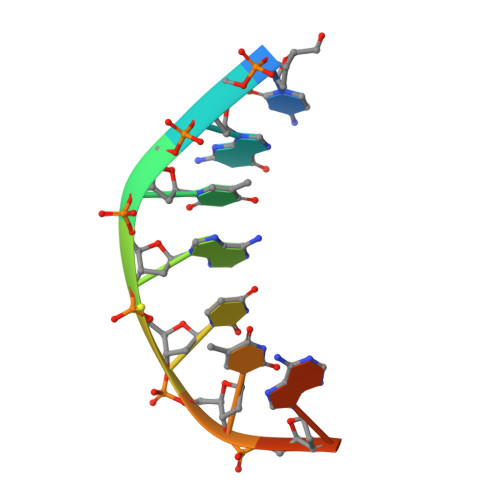
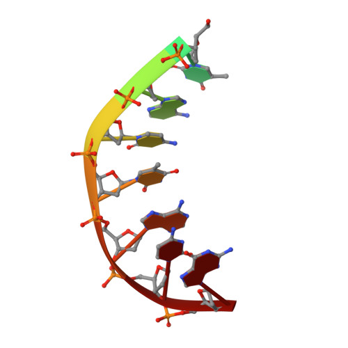
Mth0212 (WT) in complex with a 7bp dsDNA


Experimental Data Snapshot

  • Method: X-RAY DIFFRACTION
  • Resolution: 2.64 Å
  • R-Value Free: 0.297 
  • R-Value Work: 0.240 
  • R-Value Observed: 0.243 

Starting Model: experimental
View more details

wwPDB Validation   3D Report Full Report


This is version 1.4 of the entry. See complete history


Literature

Crystal Structure Analysis of DNA Uridine Endonuclease Mth212 Bound to DNA

Lakomek, K.Dickmanns, A.Ciirdaeva, E.Schomacher, L.Ficner, R.

(2010) J Mol Biol 399: 604-617

  • DOI: https://doi.org/10.1016/j.jmb.2010.04.044
  • Primary Citation of Related Structures:  
    3FZI, 3G00, 3G0A, 3G0R, 3G1K, 3G2C, 3G2D, 3G38, 3G3C, 3G3Y, 3G4T, 3G8V, 3G91, 3GA6

  • PubMed Abstract: 

    The reliable repair of pre-mutagenic U/G mismatches that originated from hydrolytic cytosine deamination is crucial for the maintenance of the correct genomic information. In most organisms, any uracil base in DNA is attacked by uracil DNA glycosylases (UDGs), but at least in Methanothermobacter thermautotrophicus DeltaH, an alternative strategy has evolved. The exonuclease III homologue Mth212 from the thermophilic archaeon M. thermautotrophicus DeltaH exhibits a DNA uridine endonuclease activity in addition to the apyrimidinic/apurinic site endonuclease and 3'-->5'exonuclease functions. Mth212 alone compensates for the lack of a UDG in a single-step reaction thus substituting the two-step pathway that requires the consecutive action of UDG and apyrimidinic/apurinic site endonuclease. In order to gain deeper insight into the structural basis required for the specific uridine recognition by Mth212, we have characterized the enzyme by means of X-ray crystallography. Structures of Mth212 wild-type or mutant proteins either alone or in complex with DNA substrates and products have been determined to a resolution of up to 1.2 A, suggesting key residues for the uridine endonuclease activity. The insertion of the side chain of Arg209 into the DNA helical base stack resembles interactions observed in human UDG and seems to be crucial for the uridine recognition. In addition, Ser171, Asn153, and Lys125 in the substrate binding pocket appear to have important functions in the discrimination of aberrant uridine against naturally occurring thymidine and cytosine residues in double-stranded DNA.


  • Organizational Affiliation

    Department of Molecular Structural Biology, Institute of Microbiology and Genetics, Georg-August University Göttingen, Justus-von-Liebig Weg 11, D-37077 Göttingen, Germany.


Macromolecules

Find similar proteins by:  (by identity cutoff)  |  3D Structure
Entity ID: 1
MoleculeChains Sequence LengthOrganismDetailsImage
Exodeoxyribonuclease
A, B
265Methanothermobacter thermautotrophicus str. Delta HMutation(s): 1 
Gene Names: mth0212MTH212MTH_212
EC: 3.1.11.2 (PDB Primary Data), 4.2.99.18 (UniProt)
UniProt
Find proteins for O26314 (Methanothermobacter thermautotrophicus (strain ATCC 29096 / DSM 1053 / JCM 10044 / NBRC 100330 / Delta H))
Explore O26314 
Go to UniProtKB:  O26314
Entity Groups  
Sequence Clusters30% Identity50% Identity70% Identity90% Identity95% Identity100% Identity
UniProt GroupO26314
Sequence Annotations
Expand
  • Reference Sequence

Find similar nucleic acids by:  Sequence   |   3D Structure  

Entity ID: 2
MoleculeChains LengthOrganismImage
5'-D(*CP*GP*TP*AP*(UPS)P*TP*AP*CP*G)-3'C [auth H]9N/A
Sequence Annotations
Expand
  • Reference Sequence

Find similar nucleic acids by:  Sequence   |   3D Structure  

Entity ID: 3
MoleculeChains LengthOrganismImage
5'-D(*CP*G*TP*AP*CP*TP*AP*CP*G)-3'D [auth I]9N/A
Sequence Annotations
Expand
  • Reference Sequence
Experimental Data & Validation

Experimental Data

  • Method: X-RAY DIFFRACTION
  • Resolution: 2.64 Å
  • R-Value Free: 0.297 
  • R-Value Work: 0.240 
  • R-Value Observed: 0.243 
  • Space Group: P 21 21 2
Unit Cell:
Length ( Å )Angle ( ˚ )
a = 100.764α = 90
b = 79.6β = 90
c = 99.409γ = 90
Software Package:
Software NamePurpose
DENZOdata reduction
SCALEPACKdata scaling
MOLREPphasing
REFMACrefinement
PDB_EXTRACTdata extraction
MAR345data collection
HKL-2000data reduction
HKL-2000data scaling

Structure Validation

View Full Validation Report


Entry History 

Deposition Data

Revision History  (Full details and data files)

  • Version 1.0: 2010-03-09
    Type: Initial release
  • Version 1.1: 2011-07-13
    Changes: Version format compliance
  • Version 1.2: 2014-02-12
    Changes: Derived calculations
  • Version 1.3: 2017-11-01
    Changes: Refinement description
  • Version 1.4: 2023-11-01
    Changes: Data collection, Database references, Derived calculations, Refinement description