3G23 | pdb_00003g23

Crystal structure of a ld-carboxypeptidase a (saro_1426) from novosphingobium aromaticivorans dsm at 1.89 A resolution


Experimental Data Snapshot

  • Method: X-RAY DIFFRACTION
  • Resolution: 1.89 Å
  • R-Value Free: 
    0.207 (Depositor), 0.209 (DCC) 
  • R-Value Work: 
    0.171 (Depositor), 0.174 (DCC) 
  • R-Value Observed: 
    0.173 (Depositor) 

wwPDB Validation 3D Report Full Report

Validation slider image for 3G23

This is version 1.6 of the entry. See complete history

Literature

Structure and function of a novel LD-carboxypeptidase a involved in peptidoglycan recycling.

Das, D.Herve, M.Elsliger, M.A.Kadam, R.U.Grant, J.C.Chiu, H.J.Knuth, M.W.Klock, H.E.Miller, M.D.Godzik, A.Lesley, S.A.Deacon, A.M.Mengin-Lecreulx, D.Wilson, I.A.

(2013) J Bacteriol 195: 5555-5566

  • DOI: https://doi.org/10.1128/JB.00900-13
  • Primary Citation Related Structures: 
    3G23

  • PubMed Abstract: 

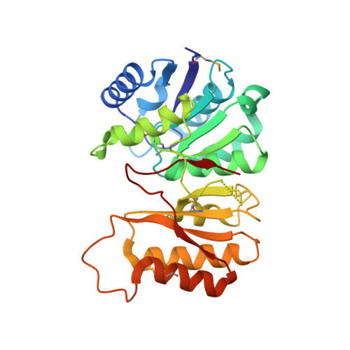
    Approximately 50% of cell wall peptidoglycan in Gram-negative bacteria is recycled with each generation. The primary substrates used for peptidoglycan biosynthesis and recycling in the cytoplasm are GlcNAc-MurNAc(anhydro)-tetrapeptide and its degradation product, the free tetrapeptide. This complex process involves ∼15 proteins, among which the cytoplasmic enzyme ld-carboxypeptidase A (LdcA) catabolizes the bond between the last two l- and d-amino acid residues in the tetrapeptide to form the tripeptide, which is then utilized as a substrate by murein peptide ligase (Mpl). LdcA has been proposed as an antibacterial target. The crystal structure of Novosphingobium aromaticivorans DSM 12444 LdcA (NaLdcA) was determined at 1.89-Å resolution. The enzyme was biochemically characterized and its interactions with the substrate modeled, identifying residues potentially involved in substrate binding. Unaccounted electron density at the dimer interface in the crystal suggested a potential site for disrupting protein-protein interactions should a dimer be required to perform its function in bacteria. Our analysis extends the identification of functional residues to several other homologs, which include enzymes from bacteria that are involved in hydrocarbon degradation and destruction of coral reefs. The NaLdcA crystal structure provides an alternate system for investigating the structure-function relationships of LdcA and increases the structural coverage of the protagonists in bacterial cell wall recycling.


  • Organizational Affiliation
    • Joint Center for Structural Genomics‡

Macromolecule Content 

  • Total Structure Weight: 61.74 kDa 
  • Atom Count: 4,681 
  • Modeled Residue Count: 540 
  • Deposited Residue Count: 548 
  • Unique protein chains: 1

Macromolecules

Find similar proteins by:|  3D Structure
Entity ID: 1
MoleculeChains  Sequence LengthOrganismDetailsImage
LD-carboxypeptidase A
A, B
274Novosphingobium aromaticivorans DSM 12444Mutation(s): 0 
Gene Names: Saro_1426YP_496704.1
UniProt
Find proteins for Q2G8F3 (Novosphingobium aromaticivorans (strain ATCC 700278 / DSM 12444 / CCUG 56034 / CIP 105152 / NBRC 16084 / F199))
Explore Q2G8F3 
Go to UniProtKB:  Q2G8F3
Entity Groups
Sequence Clusters30% Identity50% Identity70% Identity90% Identity95% Identity100% Identity
UniProt GroupQ2G8F3
Sequence Annotations
Expand
Reference Sequence

Small Molecules

Ligands 3 Unique
IDChains Name / Formula / InChI Key2D Diagram3D Interactions
SO4

Query on SO4


Download:Ideal Coordinates CCD File
C [auth A]
D [auth A]
E [auth A]
F [auth A]
G [auth A]
C [auth A],
D [auth A],
E [auth A],
F [auth A],
G [auth A],
H [auth A],
I [auth A],
J [auth A],
K [auth A],
L [auth A],
M [auth A],
N [auth A],
U [auth B],
V [auth B],
W [auth B],
X [auth B],
Y [auth B]
SULFATE ION
O4 S
QAOWNCQODCNURD-UHFFFAOYSA-L
GOL

Query on GOL


Download:Ideal Coordinates CCD File
AA [auth B]
BA [auth B]
O [auth A]
P [auth A]
Q [auth A]
AA [auth B],
BA [auth B],
O [auth A],
P [auth A],
Q [auth A],
R [auth A],
S [auth A],
Z [auth B]
GLYCEROL
C3 H8 O3
PEDCQBHIVMGVHV-UHFFFAOYSA-N
UNL

Query on UNL


Download:Ideal Coordinates CCD File
T [auth B]Unknown ligand
PEDCQBHIVMGVHV-UHFFFAOYSA-N
Modified Residues  1 Unique
IDChains TypeFormula2D DiagramParent
MSE
Query on MSE
A, B
L-PEPTIDE LINKINGC5 H11 N O2 SeMET

Experimental Data & Validation

Experimental Data

  • Method: X-RAY DIFFRACTION
  • Resolution: 1.89 Å
  • R-Value Free:  0.207 (Depositor), 0.209 (DCC) 
  • R-Value Work:  0.171 (Depositor), 0.174 (DCC) 
  • R-Value Observed: 0.173 (Depositor) 
Space Group: P 21 21 21
Unit Cell:
Length ( Å )Angle ( ˚ )
a = 43.628α = 90
b = 91.042β = 90
c = 145.744γ = 90
Software Package:
Software NamePurpose
REFMACrefinement
PHENIXrefinement
SHELXphasing
MolProbitymodel building
SCALAdata scaling
PDB_EXTRACTdata extraction
MAR345data collection
MOSFLMdata reduction
autoSHARPphasing
SHELXDphasing

Structure Validation

View Full Validation Report


Entry History 

Deposition Data

Revision History  (Full details and data files)

  • Version 1.0: 2009-02-10
    Type: Initial release
  • Version 1.1: 2011-07-13
    Changes: Advisory, Source and taxonomy, Version format compliance
  • Version 1.2: 2014-09-24
    Changes: Database references
  • Version 1.3: 2017-11-01
    Changes: Refinement description
  • Version 1.4: 2019-07-24
    Changes: Data collection, Derived calculations, Refinement description
  • Version 1.5: 2023-02-01
    Changes: Database references, Derived calculations
  • Version 1.6: 2024-11-06
    Changes: Data collection, Structure summary