


Ligand recognition by the energy sensor domain of the CcpN repressor
Chaix, D., de Guillen, K., Coq, D.L., Hoh, F., Roumestand, C., Aymerich, S., Arold, S.T., Declerck, N.To be published.
Experimental Data Snapshot
Starting Model: experimental
View more details
Entity ID: 1 | |||||
|---|---|---|---|---|---|
| Molecule | Chains | Sequence Length | Organism | Details | Image |
| YqzB protein | 159 | Bacillus subtilis | Mutation(s): 0 Gene Names: yqzB | 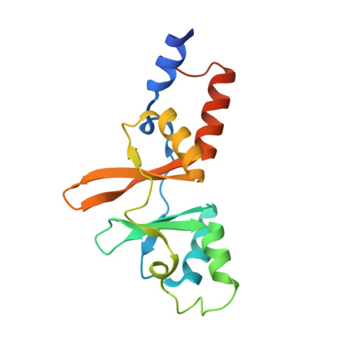 | |
UniProt | |||||
Entity Groups | |||||
| Sequence Clusters | 30% Identity50% Identity70% Identity90% Identity95% Identity100% Identity | ||||
| UniProt Group | O34994 | ||||
Sequence AnnotationsExpand | |||||
Reference Sequence | |||||
| Ligands 3 Unique | |||||
|---|---|---|---|---|---|
| ID | Chains | Name / Formula / InChI Key | 2D Diagram | 3D Interactions | |
| ANP Download:Ideal Coordinates CCD File | C [auth A], G [auth B] | PHOSPHOAMINOPHOSPHONIC ACID-ADENYLATE ESTER C10 H17 N6 O12 P3 PVKSNHVPLWYQGJ-KQYNXXCUSA-N |  | ||
| PO4 Download:Ideal Coordinates CCD File | D [auth A], H [auth B] | PHOSPHATE ION O4 P NBIIXXVUZAFLBC-UHFFFAOYSA-K |  | ||
| MG Download:Ideal Coordinates CCD File | E [auth A], F [auth A], I [auth B] | MAGNESIUM ION Mg JLVVSXFLKOJNIY-UHFFFAOYSA-N |  | ||
| Length ( Å ) | Angle ( ˚ ) |
|---|---|
| a = 54.025 | α = 90 |
| b = 103.969 | β = 90 |
| c = 98.387 | γ = 90 |
| Software Name | Purpose |
|---|---|
| SOLVE | phasing |
| REFMAC | refinement |
| MOSFLM | data reduction |
| SCALA | data scaling |


