3FWJ | pdb_00003fwj

Ferric camphor bound Cytochrome P450cam containing a selenocysteine as the 5th heme ligand, orthorombic crystal form


Experimental Data Snapshot

  • Method: X-RAY DIFFRACTION
  • Resolution: 1.90 Å
  • R-Value Free: 
    0.231 (Depositor), 0.226 (DCC) 
  • R-Value Work: 
    0.183 (Depositor), 0.180 (DCC) 
  • R-Value Observed: 
    0.185 (Depositor) 

Starting Model: experimental
View more details

wwPDB Validation 3D Report Full Report

Validation slider image for 3FWJ

Ligand Structure Quality Assessment 


This is version 1.5 of the entry. See complete history

Literature

Probing the role of the proximal heme ligand in cytochrome P450cam by recombinant incorporation of selenocysteine.

Aldag, C.Gromov, I.A.Garcia-Rubio, I.von Koenig, K.Schlichting, I.Jaun, B.Hilvert, D.

(2009) Proc Natl Acad Sci U S A 106: 5481-5486

  • DOI: https://doi.org/10.1073/pnas.0810503106
  • Primary Citation Related Structures: 
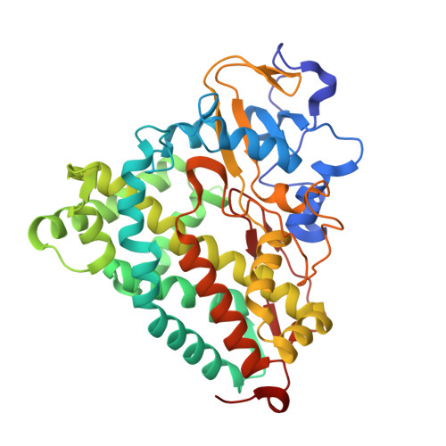
    3FWF, 3FWG, 3FWI, 3FWJ

  • PubMed Abstract: 

    The unique monooxygenase activity of cytochrome P450cam has been attributed to coordination of a cysteine thiolate to the heme cofactor. To investigate this interaction, we replaced cysteine with the more electron-donating selenocysteine. Good yields of the selenoenzyme were obtained by bacterial expression of an engineered gene containing the requisite UGA codon for selenocysteine and a simplified yet functional selenocysteine insertion sequence (SECIS). The sulfur-to-selenium substitution subtly modulates the structural, electronic, and catalytic properties of the enzyme. Catalytic activity decreases only 2-fold, whereas substrate oxidation becomes partially uncoupled from electron transfer, implying a more complex role for the axial ligand than generally assumed.


  • Organizational Affiliation
    • Laboratory of Organic Chemistry, ETH Zurich, CH-8093 Zurich, Switzerland.

Macromolecule Content 

  • Total Structure Weight: 46.45 kDa 
  • Atom Count: 3,648 
  • Modeled Residue Count: 405 
  • Deposited Residue Count: 405 
  • Unique protein chains: 1

Macromolecules

Find similar proteins by:|  3D Structure
Entity ID: 1
MoleculeChains  Sequence LengthOrganismDetailsImage
Camphor 5-monooxygenase405Pseudomonas putidaMutation(s): 3 
Gene Names: camCcyp101
EC: 1.14.15.1
UniProt
Find proteins for P00183 (Pseudomonas putida)
Explore P00183 
Go to UniProtKB:  P00183
Entity Groups
Sequence Clusters30% Identity50% Identity70% Identity90% Identity95% Identity100% Identity
UniProt GroupP00183
Sequence Annotations
Expand
Reference Sequence

Experimental Data & Validation

Experimental Data

  • Method: X-RAY DIFFRACTION
  • Resolution: 1.90 Å
  • R-Value Free:  0.231 (Depositor), 0.226 (DCC) 
  • R-Value Work:  0.183 (Depositor), 0.180 (DCC) 
  • R-Value Observed: 0.185 (Depositor) 
Space Group: P 21 21 21
Unit Cell:
Length ( Å )Angle ( ˚ )
a = 63.1α = 90
b = 65β = 90
c = 105.8γ = 90
Software Package:
Software NamePurpose
XSCALEdata scaling
REFMACrefinement
PDB_EXTRACTdata extraction
MAR345data collection
XDSdata reduction

Structure Validation

View Full Validation Report


Ligand Structure Quality Assessment 


Entry History 

Deposition Data

Revision History  (Full details and data files)

  • Version 1.0: 2009-03-03
    Type: Initial release
  • Version 1.1: 2011-07-13
    Changes: Version format compliance
  • Version 1.2: 2014-04-09
    Changes: Other
  • Version 1.3: 2014-08-06
    Changes: Other
  • Version 1.4: 2017-11-01
    Changes: Refinement description
  • Version 1.5: 2023-09-06
    Changes: Data collection, Database references, Derived calculations, Refinement description