Crystal structure of malaria parasite nucleosome assembly protein: distinct modes of protein localization and histone recognition.
Gill, J., Yogavel, M., Kumar, A., Belrhali, H., Jain, S.K., Rug, M., Brown, M., Maier, A.G., Sharma, A.(2009) J Biological Chem 284: 10076-10087
- PubMed: 19176479 Search on PubMedSearch on PubMed Central
- DOI: https://doi.org/10.1074/jbc.M808633200
- Primary Citation Related Structures:
3FS3 - PubMed Abstract:
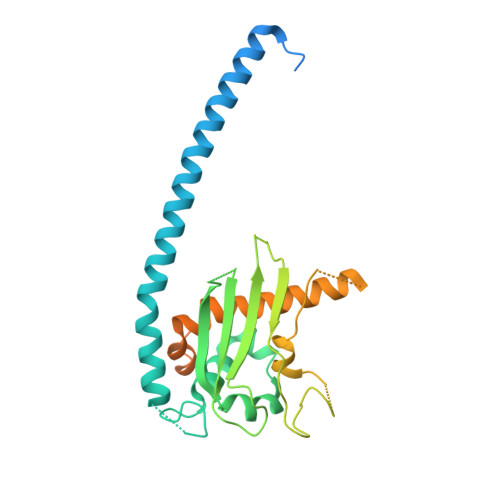
Nucleosome assembly proteins (NAPs) are histone chaperones that are essential for the transfer and incorporation of histones into nucleosomes. NAPs participate in assembly and disassembly of nucleosomes and in chromatin structure organization. Human malaria parasite Plasmodium falciparum contains two nucleosome assembly proteins termed PfNapL and PfNapS. To gain structural insights into the mechanism of NAPs, we have determined and analyzed the crystal structure of PfNapL at 2.3 A resolution. PfNapL, an ortholog of eukaryotic NAPs, is dimeric in nature and adopts a characteristic fold seen previously for yeast NAP-1 and Vps75 and for human SET/TAF-1b (beta)/INHAT. The PfNapL monomer is comprised of domain I, containing a dimerization alpha-helix, and a domain II, composed of alpha-helices and a beta-subdomain. Structural comparisons reveal that the "accessory domain," which is inserted between the domain I and domain II in yeast NAP-1 and other eukaryotic NAPs, is surprisingly absent in PfNapL. Expression of green fluorescent protein-tagged PfNapL confirmed its exclusive localization to the parasite cytoplasm. Attempts to disrupt the PfNapL gene were not successful, indicating its essential role for the malaria parasite. A detailed analysis of PfNapL structure suggests unique histone binding properties. The crucial structural differences observed between parasite and yeast NAPs shed light on possible new modes of histone recognition by nucleosome assembly proteins.
- Structural and Computational Biology Group, International Centre for Genetic Engineering and Biotechnology, Aruna Asaf Ali Road, New Delhi 110067, India.
Organizational Affiliation: