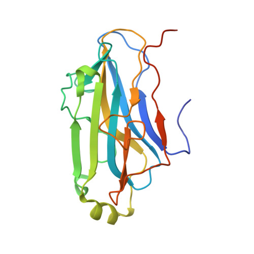
3FNK

Crystal structure of the second type II cohesin module from the cellulosomal adaptor ScaA scaffoldin of Acetivibrio cellulolyticus


Experimental Data Snapshot

  • Method: X-RAY DIFFRACTION
  • Resolution: 1.99 Å
  • R-Value Free: 0.229 
  • R-Value Work: 0.182 
  • R-Value Observed: 0.185 

Starting Model: experimental
View more details

wwPDB Validation   3D Report Full Report


This is version 1.3 of the entry. See complete history


Literature

Intermodular Linker Flexibility Revealed from Crystal Structures of Adjacent Cellulosomal Cohesins of Acetivibrio cellulolyticus

Noach, I.Frolow, F.Alber, O.Lamed, R.Shimon, L.J.W.Bayer, E.A.

(2009) J Mol Biol 391: 86-97

  • DOI: https://doi.org/10.1016/j.jmb.2009.06.006
  • Primary Citation of Related Structures:  
    1ZV9, 3BWZ, 3FNK, 3GHP

  • PubMed Abstract: 

    Cellulosome complexes comprise an intercalated set of multimodular dockerin-containing enzymatic subunits connected to cohesin-containing nonenzymatic subunits called scaffoldins. The adjoining modules in each cellulosomal subunit are interconnected by a variety of linker segments of different lengths and composition. The exact role of the cellulosomal linkers has yet to be described, although it is assumed that they contribute to the architecture and action of the cellulosome by providing the protein subunits with flexibility and by providing spacers between the enzymatic modules that could enhance interactions with the cellulose substrate. Here we present four crystal structures of Acetivibrio cellulolyticus cellulosomal type II cohesins with linker extensions. Two of the structures represent two different crystal forms (trigonal and orthorhombic) of the same N-terminal cohesin module (CohB1) together with its full (6-residue) native C-terminal linker, derived from scaffoldin B. The other two structures belong to the adjacent (second) cohesin module (CohB2), each of which was crystallized with the same 6-residue linker segment, but now positioned at the N-terminus and with either a truncated (5-residue) or a full-length (45-residue) C-terminal linker, respectively. Comparison between the two CohB1 structures revealed significant differences in the conformation of their equivalent C-terminal linker segment. In one crystal form a helical conformation was observed, as opposed to an extended conformation in the other. The CohB2 structures also displayed diverse conformations in their linker segments. In these structures, different linker conformations were observed in the individual molecules within the asymmetric unit of each structure. This conformational diversity implies that the linkers may adopt alternative conformations in their natural environment, consistent with varying environmental conditions. The findings suggest that linkers can play an important role in the assembly, dynamics and function of the cellulosomal components.


  • Organizational Affiliation

    Department of Biological Chemistry, The Weizmann Institute of Science, Rehovot, Israel.


Macromolecules
Find similar proteins by:  (by identity cutoff)  |  3D Structure
Entity ID: 1
MoleculeChains Sequence LengthOrganismDetailsImage
Cellulosomal scaffoldin adaptor protein B
A, B, C
187Acetivibrio cellulolyticusMutation(s): 0 
Gene Names: scaB
UniProt
Find proteins for Q7WYN3 (Acetivibrio cellulolyticus)
Explore Q7WYN3 
Go to UniProtKB:  Q7WYN3
Entity Groups  
Sequence Clusters30% Identity50% Identity70% Identity90% Identity95% Identity100% Identity
UniProt GroupQ7WYN3
Sequence Annotations
Expand
  • Reference Sequence
Small Molecules
Ligands 4 Unique
IDChains Name / Formula / InChI Key2D Diagram3D Interactions
BU1
Query on BU1

Download Ideal Coordinates CCD File 
L [auth B]1,4-BUTANEDIOL
C4 H10 O2
WERYXYBDKMZEQL-UHFFFAOYSA-N
PDO
Query on PDO

Download Ideal Coordinates CCD File 
F [auth A],
N [auth B],
S [auth C]
1,3-PROPANDIOL
C3 H8 O2
YPFDHNVEDLHUCE-UHFFFAOYSA-N
EDO
Query on EDO

Download Ideal Coordinates CCD File 
D [auth A]
G [auth B]
H [auth B]
I [auth B]
J [auth B]
D [auth A],
G [auth B],
H [auth B],
I [auth B],
J [auth B],
K [auth B],
Q [auth C],
R [auth C]
1,2-ETHANEDIOL
C2 H6 O2
LYCAIKOWRPUZTN-UHFFFAOYSA-N
ACT
Query on ACT

Download Ideal Coordinates CCD File 
E [auth A],
M [auth B],
O [auth C],
P [auth C]
ACETATE ION
C2 H3 O2
QTBSBXVTEAMEQO-UHFFFAOYSA-M
Experimental Data & Validation

Experimental Data

  • Method: X-RAY DIFFRACTION
  • Resolution: 1.99 Å
  • R-Value Free: 0.229 
  • R-Value Work: 0.182 
  • R-Value Observed: 0.185 
  • Space Group: P 21 21 21
Unit Cell:
Length ( Å )Angle ( ˚ )
a = 90.359α = 90
b = 68.645β = 90
c = 111.294γ = 90
Software Package:
Software NamePurpose
ProDCdata collection
MOLREPphasing
REFMACrefinement
HKL-2000data reduction
SCALEPACKdata scaling

Structure Validation

View Full Validation Report


Entry History 

Deposition Data

Revision History  (Full details and data files)

  • Version 1.0: 2009-06-23
    Type: Initial release
  • Version 1.1: 2011-07-13
    Changes: Version format compliance
  • Version 1.2: 2017-11-01
    Changes: Refinement description
  • Version 1.3: 2023-11-01
    Changes: Data collection, Database references, Derived calculations, Refinement description