3FFD | pdb_00003ffd

Structure of parathyroid hormone-related protein complexed to a neutralizing monoclonal antibody


Experimental Data Snapshot

  • Method: X-RAY DIFFRACTION
  • Resolution: 2.00 Å
  • R-Value Free: 
    0.271 (Depositor) 
  • R-Value Work: 
    0.224 (Depositor), 0.240 (DCC) 
  • R-Value Observed: 
    0.224 (Depositor) 

Starting Models: experimental
View more details

wwPDB Validation 3D Report Full Report

Validation slider image for 3FFD

This is version 1.4 of the entry. See complete history

Literature

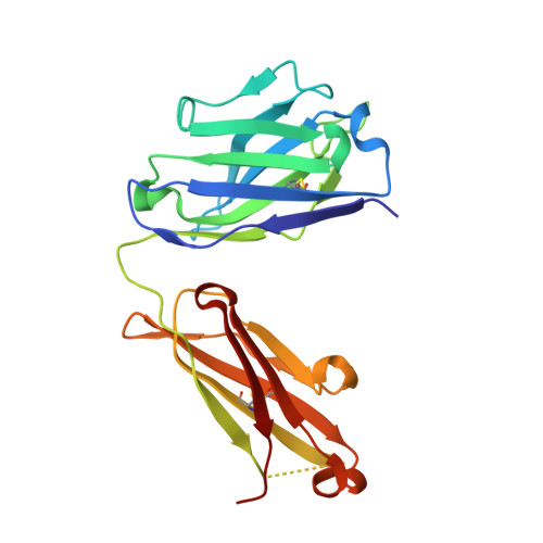
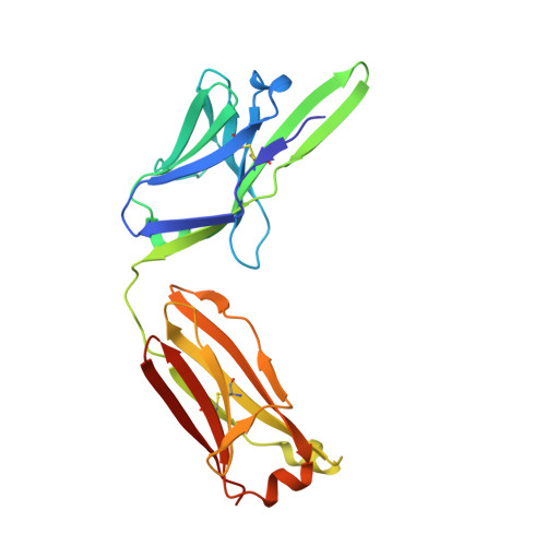
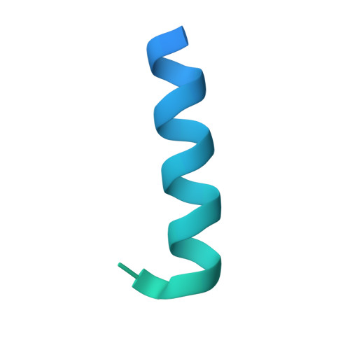
Structural basis for antibody discrimination between two hormones that recognize the parathyroid hormone receptor

McKinstry, W.J.Polekhina, G.Diefenbach-Jagger, H.Ho, P.W.M.Sato, K.Onuma, E.Gillespie, M.T.Martin, T.J.Parker, M.W.

(2009) J Biological Chem 284: 15557-15563

  • DOI: https://doi.org/10.1074/jbc.M900044200
  • Primary Citation Related Structures: 
    3FFD

  • PubMed Abstract: 

    Parathyroid hormone-related protein (PTHrP) plays a vital role in the embryonic development of the skeleton and other tissues. When it is produced in excess by cancers it can cause hypercalcemia, and its local production by breast cancer cells has been implicated in the pathogenesis of bone metastasis formation in that disease. Antibodies have been developed that neutralize the action of PTHrP through its receptor, parathyroid hormone receptor 1, without influencing parathyroid hormone action through the same receptor. Such neutralizing antibodies against PTHrP are therapeutically effective in animal models of the humoral hypercalcemia of malignancy and of bone metastasis formation. We have determined the crystal structure of the complex between PTHrP (residues 1-108) and a neutralizing monoclonal anti-PTHrP antibody that reveals the only point of contact is an alpha-helical structure extending from residues 14-29. Another striking feature is that the same residues that interact with the antibody also interact with parathyroid hormone receptor 1, showing that the antibody and the receptor binding site on the hormone closely overlap. The structure explains how the antibody discriminates between the two hormones and provides information that could be used in the development of novel agonists and antagonists of their common receptor.


  • Organizational Affiliation
    • Biota Structural Biology Laboratory, St. Vincent's Institute of Medical Research, Fitzroy, Victoria 3065, Australia.

Macromolecule Content 

  • Total Structure Weight: 59.69 kDa 
  • Atom Count: 3,595 
  • Modeled Residue Count: 445 
  • Deposited Residue Count: 546 
  • Unique protein chains: 3

Macromolecules

Find similar proteins by:|  3D Structure
Entity ID: 1
MoleculeChains  Sequence LengthOrganismDetailsImage
Monoclonal antibody, heavy chain, Fab fragment218Mus musculusMutation(s): 0 
Find similar proteins by:|  3D Structure
Entity ID: 2
MoleculeChains  Sequence LengthOrganismDetailsImage
Monoclonal antibody, light chain, Fab fragment220Mus musculusMutation(s): 0 
Find similar proteins by:|  3D Structure
Entity ID: 3
MoleculeChains  Sequence LengthOrganismDetailsImage
Parathyroid hormone-related proteinC [auth P]108Homo sapiensMutation(s): 0 
Membrane Entity: Yes 
UniProt & NIH Common Fund Data Resources
Find proteins for P12272 (Homo sapiens)
Explore P12272 
Go to UniProtKB:  P12272
PHAROS:  P12272
GTEx:  ENSG00000087494 
Entity Groups
Sequence Clusters30% Identity50% Identity70% Identity90% Identity95% Identity100% Identity
UniProt GroupP12272
Sequence Annotations
Expand
Reference Sequence

Experimental Data & Validation

Experimental Data

  • Method: X-RAY DIFFRACTION
  • Resolution: 2.00 Å
  • R-Value Free:  0.271 (Depositor) 
  • R-Value Work:  0.224 (Depositor), 0.240 (DCC) 
  • R-Value Observed: 0.224 (Depositor) 
Space Group: P 21 21 2
Unit Cell:
Length ( Å )Angle ( ˚ )
a = 72.613α = 90
b = 96.285β = 90
c = 88.544γ = 90
Software Package:
Software NamePurpose
ADSCdata collection
AMoREphasing
CNSrefinement
DENZOdata reduction
SCALEPACKdata scaling

Structure Validation

View Full Validation Report


Entry History 

Deposition Data

Revision History  (Full details and data files)

  • Version 1.0: 2009-04-28
    Type: Initial release
  • Version 1.1: 2011-07-13
    Changes: Version format compliance
  • Version 1.2: 2014-02-05
    Changes: Database references
  • Version 1.3: 2023-11-01
    Changes: Data collection, Database references, Refinement description
  • Version 1.4: 2024-11-13
    Changes: Structure summary