3FF8

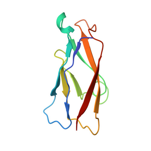
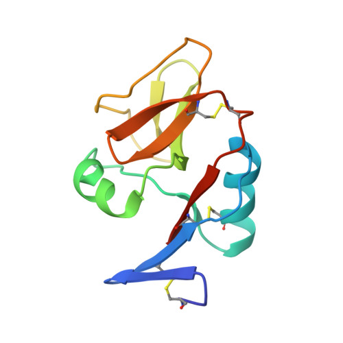
Structure of NK cell receptor KLRG1 bound to E-cadherin


Experimental Data Snapshot

  • Method: X-RAY DIFFRACTION
  • Resolution: 2.00 Å
  • R-Value Free: 0.253 
  • R-Value Work: 0.212 

Starting Models: experimental
View more details

wwPDB Validation   3D Report Full Report


This is version 1.4 of the entry. See complete history


Literature

Structure of natural killer cell receptor KLRG1 bound to E-cadherin reveals basis for MHC-independent missing self recognition.

Li, Y.Hofmann, M.Wang, Q.Teng, L.Chlewicki, L.K.Pircher, H.Mariuzza, R.A.

(2009) Immunity 31: 35-46

  • DOI: https://doi.org/10.1016/j.immuni.2009.04.019
  • Primary Citation of Related Structures:  
    3FF7, 3FF8, 3FF9

  • PubMed Abstract: 

    The cytolytic activity of natural killer (NK) cells is regulated by inhibitory receptors that detect the absence of self molecules on target cells. Structural studies of missing self recognition have focused on NK receptors that bind MHC. However, NK cells also possess inhibitory receptors specific for non-MHC ligands, notably cadherins, which are downregulated in metastatic tumors. We determined the structure of killer cell lectin-like receptor G1 (KLRG1) in complex with E-cadherin. KLRG1 mediates missing self recognition by binding to a highly conserved site on classical cadherins, enabling it to monitor expression of several cadherins (E-, N-, and R-) on target cells. This site overlaps the site responsible for cell-cell adhesion but is distinct from the integrin alpha(E)beta(7) binding site. We propose that E-cadherin may coengage KLRG1 and alpha(E)beta(7) and that KLRG1 overcomes its exceptionally weak affinity for cadherins through multipoint attachment to target cells, resulting in inhibitory signaling.


  • Organizational Affiliation

    Center for Advanced Research in Biotechnology, WM Keck Laboratory for Structural Biology, University of Maryland Biotechnology Institute, Rockville, MD 20850, USA.


Macromolecules
Find similar proteins by:  (by identity cutoff)  |  3D Structure
Entity ID: 1
MoleculeChains Sequence LengthOrganismDetailsImage
Epithelial cadherin
A, B
101Homo sapiensMutation(s): 1 
Gene Names: CDH1CDHEUVO
UniProt & NIH Common Fund Data Resources
Find proteins for P12830 (Homo sapiens)
Explore P12830 
Go to UniProtKB:  P12830
PHAROS:  P12830
GTEx:  ENSG00000039068 
Entity Groups  
Sequence Clusters30% Identity50% Identity70% Identity90% Identity95% Identity100% Identity
UniProt GroupP12830
Sequence Annotations
Expand
  • Reference Sequence
Find similar proteins by:  (by identity cutoff)  |  3D Structure
Entity ID: 2
MoleculeChains Sequence LengthOrganismDetailsImage
Killer cell lectin-like receptor subfamily G member 1
C, D
114Mus musculusMutation(s): 0 
Gene Names: Klrg1Mafa
UniProt
Find proteins for O88713 (Mus musculus)
Explore O88713 
Go to UniProtKB:  O88713
Entity Groups  
Sequence Clusters30% Identity50% Identity70% Identity90% Identity95% Identity100% Identity
UniProt GroupO88713
Sequence Annotations
Expand
  • Reference Sequence
Experimental Data & Validation

Experimental Data

  • Method: X-RAY DIFFRACTION
  • Resolution: 2.00 Å
  • R-Value Free: 0.253 
  • R-Value Work: 0.212 
  • Space Group: P 21 21 21
Unit Cell:
Length ( Å )Angle ( ˚ )
a = 53.776α = 90
b = 90.526β = 90
c = 107.593γ = 90
Software Package:
Software NamePurpose
DENZOdata reduction
SCALEPACKdata scaling
PHASERphasing
CNSrefinement
PDB_EXTRACTdata extraction

Structure Validation

View Full Validation Report


Entry History 

Deposition Data

Revision History  (Full details and data files)

  • Version 1.0: 2009-07-28
    Type: Initial release
  • Version 1.1: 2011-07-13
    Changes: Version format compliance
  • Version 1.2: 2017-11-01
    Changes: Advisory, Refinement description
  • Version 1.3: 2021-10-20
    Changes: Advisory, Database references, Derived calculations
  • Version 1.4: 2023-09-06
    Changes: Data collection, Refinement description