Tuning a three-component reaction for trapping kinase substrate complexes.
Statsuk, A.V., Maly, D.J., Seeliger, M.A., Fabian, M.A., Biggs, W.H., Lockhart, D.J., Zarrinkar, P.P., Kuriyan, J., Shokat, K.M.(2008) J Am Chem Soc 130: 17568-17574
- PubMed: 19053485 Search on PubMedSearch on PubMed Central
- DOI: https://doi.org/10.1021/ja807066f
- Primary Citation Related Structures:
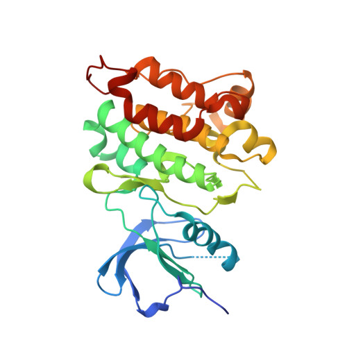
3F6X - PubMed Abstract:
The upstream protein kinases responsible for thousands of phosphorylation events in the phosphoproteome remain to be discovered. We developed a three-component chemical reaction which converts the transient noncovalent substrate-kinase complex into a covalently cross-linked product by utilizing a dialdehyde-based cross-linker, 1. Unfortunately, the reaction of 1 with a lysine in the kinase active site and an engineered cysteine on the substrate to form an isoindole cross-linked product could not be performed in the presence of competing cellular proteins due to nonspecific side reactions. In order to more selectively target the cross-linker to protein kinases in cell lysates, we replaced the weak, kinase-binding adenosine moiety of 1 with a potent protein kinase inhibitor scaffold. In addition, we replaced the o-phthaldialdehyde moiety in 1 with a less-reactive thiophene-2,3-dicarboxaldehyde moiety. The combination of these two structural modifications provides for cross-linking of a cysteine-containing substrate to its corresponding kinase in the presence of competing cellular proteins.
- Howard Hughes Medical Institute, Department of Cellular and Molecular Pharmacology, University of California, San Francisco, California 94107, USA.
Organizational Affiliation: