3F37 | pdb_00003f37

Apoferritin: complex with 2,6-dimethylphenol


Experimental Data Snapshot

  • Method: X-RAY DIFFRACTION
  • Resolution: 1.54 Å
  • R-Value Free: 
    0.198 (Depositor), 0.200 (DCC) 
  • R-Value Work: 
    0.174 (Depositor), 0.176 (DCC) 
  • R-Value Observed: 
    0.175 (Depositor) 

Starting Model: experimental
View more details

wwPDB Validation 3D Report Full Report

Validation slider image for 3F37

This is version 1.2 of the entry. See complete history

Literature

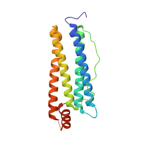
A unitary anesthetic binding site at high resolution.

Vedula, L.S.Brannigan, G.Economou, N.J.Xi, J.Hall, M.A.Liu, R.Rossi, M.J.Dailey, W.P.Grasty, K.C.Klein, M.L.Eckenhoff, R.G.Loll, P.J.

(2009) J Biological Chem 284: 24176-24184

  • DOI: https://doi.org/10.1074/jbc.M109.017814
  • Primary Citation Related Structures: 
    3F32, 3F33, 3F34, 3F35, 3F36, 3F37, 3F38, 3F39

  • PubMed Abstract: 

    Propofol is the most widely used injectable general anesthetic. Its targets include ligand-gated ion channels such as the GABA(A) receptor, but such receptor-channel complexes remain challenging to study at atomic resolution. Until structural biology methods advance to the point of being able to deal with systems such as the GABA(A) receptor, it will be necessary to use more tractable surrogates to probe the molecular details of anesthetic recognition. We have previously shown that recognition of inhalational general anesthetics by the model protein apoferritin closely mirrors recognition by more complex and clinically relevant protein targets; here we show that apoferritin also binds propofol and related GABAergic anesthetics, and that the same binding site mediates recognition of both inhalational and injectable anesthetics. Apoferritin binding affinities for a series of propofol analogs were found to be strongly correlated with the ability to potentiate GABA responses at GABA(A) receptors, validating this model system for injectable anesthetics. High resolution x-ray crystal structures reveal that, despite the presence of hydrogen bond donors and acceptors, anesthetic recognition is mediated largely by van der Waals forces and the hydrophobic effect. Molecular dynamics simulations indicate that the ligands undergo considerable fluctuations about their equilibrium positions. Finally, apoferritin displays both structural and dynamic responses to anesthetic binding, which may mimic changes elicited by anesthetics in physiologic targets like ion channels.


  • Organizational Affiliation
    • Department of Anesthesiology and Critical Care, University of Pennsylvania, Philadelphia, Pennsylvania 19104, USA.

Macromolecule Content 

  • Total Structure Weight: 21.13 kDa 
  • Atom Count: 1,658 
  • Modeled Residue Count: 168 
  • Deposited Residue Count: 174 
  • Unique protein chains: 1

Macromolecules

Find similar proteins by:|  3D Structure
Entity ID: 1
MoleculeChains  Sequence LengthOrganismDetailsImage
Ferritin light chain174Equus caballusMutation(s): 0 
UniProt
Find proteins for P02791 (Equus caballus)
Explore P02791 
Go to UniProtKB:  P02791
Entity Groups
Sequence Clusters30% Identity50% Identity70% Identity90% Identity95% Identity100% Identity
UniProt GroupP02791
Sequence Annotations
Expand
Reference Sequence

Small Molecules

Ligands 4 Unique
IDChains Name / Formula / InChI Key2D Diagram3D Interactions
2MY

Query on 2MY


Download:Ideal Coordinates CCD File
I [auth A]2,6-dimethylphenol
C8 H10 O
NXXYKOUNUYWIHA-UHFFFAOYSA-N
CD

Query on CD


Download:Ideal Coordinates CCD File
B [auth A]
C [auth A]
D [auth A]
E [auth A]
F [auth A]
B [auth A],
C [auth A],
D [auth A],
E [auth A],
F [auth A],
G [auth A],
H [auth A]
CADMIUM ION
Cd
WLZRMCYVCSSEQC-UHFFFAOYSA-N
SO4

Query on SO4


Download:Ideal Coordinates CCD File
J [auth A],
K [auth A],
L [auth A]
SULFATE ION
O4 S
QAOWNCQODCNURD-UHFFFAOYSA-L
ACT

Query on ACT


Download:Ideal Coordinates CCD File
M [auth A]ACETATE ION
C2 H3 O2
QTBSBXVTEAMEQO-UHFFFAOYSA-M

Experimental Data & Validation

Experimental Data

  • Method: X-RAY DIFFRACTION
  • Resolution: 1.54 Å
  • R-Value Free:  0.198 (Depositor), 0.200 (DCC) 
  • R-Value Work:  0.174 (Depositor), 0.176 (DCC) 
  • R-Value Observed: 0.175 (Depositor) 
Space Group: F 4 3 2
Unit Cell:
Length ( Å )Angle ( ˚ )
a = 181.252α = 90
b = 181.252β = 90
c = 181.252γ = 90
Software Package:
Software NamePurpose
REFMACrefinement
d*TREKdata reduction
d*TREKdata scaling
REFMACphasing

Structure Validation

View Full Validation Report


Entry History 

Deposition Data

Revision History  (Full details and data files)

  • Version 1.0: 2009-07-14
    Type: Initial release
  • Version 1.1: 2011-07-13
    Changes: Version format compliance
  • Version 1.2: 2023-09-06
    Changes: Data collection, Database references, Derived calculations, Refinement description