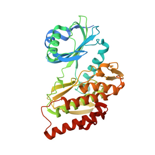
Crystal Structure of Human Haspin with an Imidazo-pyridazine ligand
Filippakopoulos, P., Eswaran, J., Keates, T., Burgess-Brown, N., Fedorov, O., Yue, W.W., Murray, J.W., Pike, A.C.W., Von Delft, F., Arrowsmith, C.H., Edwards, A.M., Weigelt, J., Bountra, C., Knapp, S.To be published.