Hit to lead account of the discovery of a new class of inhibitors of Pim kinases and crystallographic studies revealing an unusual kinase binding mode.
Qian, K., Wang, L., Cywin, C.L., Farmer, B.T., Hickey, E., Homon, C., Jakes, S., Kashem, M.A., Lee, G., Leonard, S., Li, J., Magboo, R., Mao, W., Pack, E., Peng, C., Prokopowicz, A., Welzel, M., Wolak, J., Morwick, T.(2009) J Med Chem 52: 1814-1827
- PubMed: 19256503 Search on PubMed
- DOI: https://doi.org/10.1021/jm801242y
- Primary Citation Related Structures:
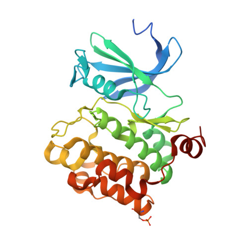
3F2A - PubMed Abstract:
A series of inhibitors of Pim-2 kinase identified by high-throughput screening is described. Details of the hit validation and lead generation process and structure-activity relationship (SAR) studies are presented. Disclosure of an unconventional binding mode for 1, as revealed by X-ray crystallography using the highly homologous Pim-1 protein, is also presented, and observed binding features are shown to correlate with the Pim-2 SAR. While highly selective within the kinase family, the series shows similar potency for both Pim-1 and Pim-2, which was expected on the basis of homology, but unusual in light of reports in the literature documenting a bias for Pim-1. A rationale for these observations based on Pim-1 and Pim-2 K(M(ATP)) values is suggested. Some interesting cross reactivity with casein kinase-2 was also identified, and structural features which may contribute to the association are discussed.
- Boehringer Ingelheim Pharmaceuticals, Inc, Ridgefield, Connecticut 06801-0368, USA.
Organizational Affiliation: