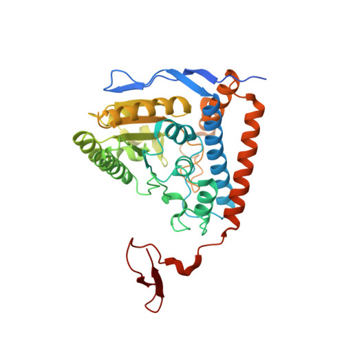
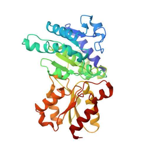
3EXE

Crystal structure of the pyruvate dehydrogenase (E1p) component of human pyruvate dehydrogenase complex


Experimental Data Snapshot

  • Method: X-RAY DIFFRACTION
  • Resolution: 1.98 Å
  • R-Value Free: 0.206 
  • R-Value Work: 0.158 
  • R-Value Observed: 0.161 

wwPDB Validation   3D Report Full Report


Ligand Structure Quality Assessment 


This is version 1.3 of the entry. See complete history


Literature

Structural basis for inactivation of the human pyruvate dehydrogenase complex by phosphorylation: role of disordered phosphorylation loops.

Kato, M.Wynn, R.M.Chuang, J.L.Tso, S.C.Machius, M.Li, J.Chuang, D.T.

(2008) Structure 16: 1849-1859

  • DOI: https://doi.org/10.1016/j.str.2008.10.010
  • Primary Citation of Related Structures:  
    3EXE, 3EXF, 3EXG, 3EXH, 3EXI

  • PubMed Abstract: 

    We report the crystal structures of the phosporylated pyruvate dehydrogenase (E1p) component of the human pyruvate dehydrogenase complex (PDC). The complete phosphorylation at Ser264-alpha (site 1) of a variant E1p protein was achieved using robust pyruvate dehydrogenase kinase 4 free of the PDC core. We show that unlike its unmodified counterpart, the presence of a phosphoryl group at Ser264-alpha prevents the cofactor thiamine diphosphate-induced ordering of the two loops carrying the three phosphorylation sites. The disordering of these phosphorylation loops is caused by a previously unrecognized steric clash between the phosphoryl group at site 1 and a nearby Ser266-alpha, which nullifies a hydrogen-bonding network essential for maintaining the loop conformations. The disordered phosphorylation loops impede the binding of lipoyl domains of the PDC core to E1p, negating the reductive acetylation step. This results in the disruption of the substrate channeling in the PDC, leading to the inactivation of this catalytic machine.


  • Organizational Affiliation

    Department of Internal Medicine, University of Texas Southwestern Medical Center, Dallas, TX 75390-9038, USA.


Macromolecules
Find similar proteins by:  (by identity cutoff)  |  3D Structure
Entity ID: 1
MoleculeChains Sequence LengthOrganismDetailsImage
Pyruvate dehydrogenase E1 component subunit alpha, somatic form, mitochondrial
A, C, E, G
382Homo sapiensMutation(s): 0 
Gene Names: PDHA1PHE1A
EC: 1.2.4.1
UniProt & NIH Common Fund Data Resources
Find proteins for P08559 (Homo sapiens)
Explore P08559 
Go to UniProtKB:  P08559
PHAROS:  P08559
GTEx:  ENSG00000131828 
Entity Groups  
Sequence Clusters30% Identity50% Identity70% Identity90% Identity95% Identity100% Identity
UniProt GroupP08559
Sequence Annotations
Expand
  • Reference Sequence
Find similar proteins by:  (by identity cutoff)  |  3D Structure
Entity ID: 2
MoleculeChains Sequence LengthOrganismDetailsImage
Pyruvate dehydrogenase E1 component subunit beta, mitochondrial
B, D, F, H
329Homo sapiensMutation(s): 0 
Gene Names: PDHBPHE1B
EC: 1.2.4.1
UniProt & NIH Common Fund Data Resources
Find proteins for P11177 (Homo sapiens)
Explore P11177 
Go to UniProtKB:  P11177
PHAROS:  P11177
GTEx:  ENSG00000168291 
Entity Groups  
Sequence Clusters30% Identity50% Identity70% Identity90% Identity95% Identity100% Identity
UniProt GroupP11177
Sequence Annotations
Expand
  • Reference Sequence
Small Molecules
Ligands 4 Unique
IDChains Name / Formula / InChI Key2D Diagram3D Interactions
TPP
Query on TPP

Download Ideal Coordinates CCD File 
J [auth A],
N [auth C],
R [auth E],
V [auth G]
THIAMINE DIPHOSPHATE
C12 H19 N4 O7 P2 S
AYEKOFBPNLCAJY-UHFFFAOYSA-O
GOL
Query on GOL

Download Ideal Coordinates CCD File 
K [auth A],
O [auth C],
S [auth E],
W [auth G]
GLYCEROL
C3 H8 O3
PEDCQBHIVMGVHV-UHFFFAOYSA-N
MN
Query on MN

Download Ideal Coordinates CCD File 
I [auth A],
M [auth C],
Q [auth E],
U [auth G]
MANGANESE (II) ION
Mn
WAEMQWOKJMHJLA-UHFFFAOYSA-N
K
Query on K

Download Ideal Coordinates CCD File 
L [auth B],
P [auth D],
T [auth F],
X [auth H]
POTASSIUM ION
K
NPYPAHLBTDXSSS-UHFFFAOYSA-N
Binding Affinity Annotations 
IDSourceBinding Affinity
TPP PDBBind:  3EXE Kd: 110 (nM) from 1 assay(s)
Experimental Data & Validation

Experimental Data

  • Method: X-RAY DIFFRACTION
  • Resolution: 1.98 Å
  • R-Value Free: 0.206 
  • R-Value Work: 0.158 
  • R-Value Observed: 0.161 
  • Space Group: P 1 21 1
Unit Cell:
Length ( Å )Angle ( ˚ )
a = 103.572α = 90
b = 129.617β = 92.48
c = 124.303γ = 90
Software Package:
Software NamePurpose
DENZOdata reduction
SCALEPACKdata scaling
REFMACrefinement
PDB_EXTRACTdata extraction
ADSCdata collection
HKL-2000data reduction

Structure Validation

View Full Validation Report


Ligand Structure Quality Assessment 


Entry History 

Deposition Data

Revision History  (Full details and data files)

  • Version 1.0: 2008-11-25
    Type: Initial release
  • Version 1.1: 2011-07-13
    Changes: Advisory, Version format compliance
  • Version 1.2: 2017-10-25
    Changes: Refinement description
  • Version 1.3: 2023-12-27
    Changes: Data collection, Database references, Derived calculations, Refinement description