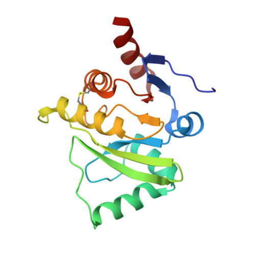
3ETI

Structure of a cubic crystal form of X (ADRP) domain from FCoV


Experimental Data Snapshot

  • Method: X-RAY DIFFRACTION
  • Resolution: 2.20 Å
  • R-Value Free: 0.181 
  • R-Value Work: 0.147 
  • R-Value Observed: 0.149 

wwPDB Validation   3D Report Full Report


This is version 1.4 of the entry. See complete history


Literature

Structure of the X (ADRP) domain of nsp3 from feline coronavirus

Wojdyla, J.A.Manolaridis, I.Snijder, E.J.Gorbalenya, A.E.Coutard, B.Piotrowski, Y.Hilgenfeld, R.Tucker, P.A.

(2009) Acta Crystallogr D Biol Crystallogr 65: 1292-1300

  • DOI: https://doi.org/10.1107/S0907444909040074
  • Primary Citation of Related Structures:  
    3ETI, 3EW5, 3JZT

  • PubMed Abstract: 

    The structure of the X (or ADRP) domain of a pathogenic variant of feline coronavirus (FCoV) has been determined in tetragonal and cubic crystal forms to 3.1 and 2.2 A resolution, respectively. In the tetragonal crystal form, glycerol-3-phosphate was observed in the ADP-ribose-binding site. Both crystal forms contained large solvent channels and had a solvent content of higher than 70%. Only very weak binding of this domain to ADP-ribose was detected in vitro. However, the structure with ADP-ribose bound was determined in the cubic crystal form at 3.9 A resolution. The structure of the FCoV X domain had the expected macro-domain fold and is the first structure of this domain from a coronavirus belonging to subgroup 1a.


  • Organizational Affiliation

    EMBL Hamburg Outstation, c/o DESY, D-22603 Hamburg, Germany.


Macromolecules
Find similar proteins by:  (by identity cutoff)  |  3D Structure
Entity ID: 1
MoleculeChains Sequence LengthOrganismDetailsImage
macro domain of Non-structural protein 3
A, B, C, D, E
A, B, C, D, E, F, G, H
168Feline infectious peritonitis virus (strain 79-1146)Mutation(s): 1 
UniProt
Find proteins for Q98VG9 (Feline coronavirus (strain FIPV WSU-79/1146))
Explore Q98VG9 
Go to UniProtKB:  Q98VG9
Entity Groups  
Sequence Clusters30% Identity50% Identity70% Identity90% Identity95% Identity100% Identity
UniProt GroupQ98VG9
Sequence Annotations
Expand
  • Reference Sequence
Experimental Data & Validation

Experimental Data

  • Method: X-RAY DIFFRACTION
  • Resolution: 2.20 Å
  • R-Value Free: 0.181 
  • R-Value Work: 0.147 
  • R-Value Observed: 0.149 
  • Space Group: P 21 3
Unit Cell:
Length ( Å )Angle ( ˚ )
a = 218.99α = 90
b = 218.99β = 90
c = 218.99γ = 90
Software Package:
Software NamePurpose
XSCALEdata scaling
PHASERphasing
REFMACrefinement
PDB_EXTRACTdata extraction
XDSdata scaling
XDSdata reduction
SCALAdata scaling

Structure Validation

View Full Validation Report


Entry History 

Deposition Data

Revision History  (Full details and data files)

  • Version 1.0: 2009-10-13
    Type: Initial release
  • Version 1.1: 2011-07-13
    Changes: Version format compliance
  • Version 1.2: 2021-11-10
    Changes: Database references
  • Version 1.3: 2023-12-27
    Changes: Data collection, Refinement description
  • Version 1.4: 2024-11-06
    Changes: Structure summary