3EQA | pdb_00003eqa

Catalytic domain of glucoamylase from aspergillus niger complexed with tris and glycerol


Experimental Data Snapshot

  • Method: X-RAY DIFFRACTION
  • Resolution: 1.90 Å
  • R-Value Free: 
    0.229 (Depositor), 0.234 (DCC) 
  • R-Value Work: 
    0.181 (Depositor), 0.197 (DCC) 
  • R-Value Observed: 
    0.184 (Depositor) 

Starting Model: experimental
View more details

wwPDB Validation 3D Report Full Report

Validation slider image for 3EQA

Ligand Structure Quality Assessment 


This is version 2.2 of the entry. See complete history

Literature

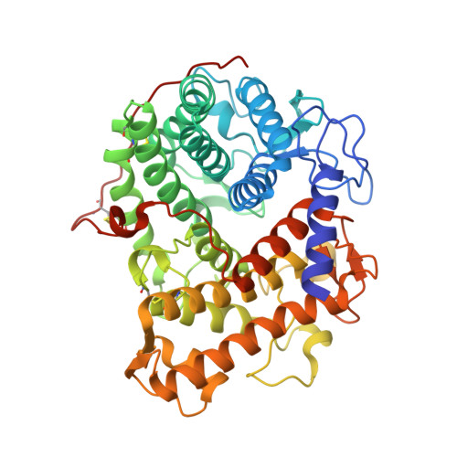
Structure of the catalytic domain of glucoamylase from Aspergillus niger.

Lee, J.Paetzel, M.

(2011) Acta Crystallogr Sect F Struct Biol Cryst Commun 67: 188-192

  • DOI: https://doi.org/10.1107/S1744309110049390
  • Primary Citation Related Structures: 
    3EQA

  • PubMed Abstract: 

    Glucoamylase from Aspergillus niger is an industrially important biocatalyst that is utilized in the mass production of glucose from raw starch or soluble oligosaccharides. The G1 isoform consists of a catalytic domain and a starch-binding domain connected by a heavily glycosylated linker region. The amino-terminal catalytic domain of the G1 isoform generated by subtilisin cleavage has been crystallized at pH 8.5, which is a significantly higher pH condition than used for previously characterized glucoamylase crystals. The refined structure at 1.9 Å resolution reveals the active site of the enzyme in complex with both Tris and glycerol molecules. The ligands display both unique and analogous interactions with the substrate-binding site when compared with previous structures of homologous enzymes bound to inhibitors.


  • Organizational Affiliation
    • Department of Molecular Biology and Biochemistry, Simon Fraser University, Burnaby, British Columbia, Canada.

Macromolecule Content 

  • Total Structure Weight: 54.12 kDa 
  • Atom Count: 4,107 
  • Modeled Residue Count: 458 
  • Deposited Residue Count: 470 
  • Unique protein chains: 1

Macromolecules

Find similar proteins by:|  3D Structure
Entity ID: 1
MoleculeChains  Sequence LengthOrganismDetailsImage
Glucoamylase470Aspergillus nigerMutation(s): 0 
EC: 3.2.1.3
UniProt
Find proteins for P69328 (Aspergillus niger)
Explore P69328 
Go to UniProtKB:  P69328
Entity Groups
Sequence Clusters30% Identity50% Identity70% Identity90% Identity95% Identity100% Identity
UniProt GroupP69328
Glycosylation
Glycosylation Sites: 9
Sequence Annotations
Expand
Reference Sequence

Oligosaccharides

Help  
Entity ID: 2
MoleculeChains Length2D Diagram GlycosylationD Interactions
alpha-D-mannopyranose-(1-3)-beta-D-mannopyranose-(1-4)-2-acetamido-2-deoxy-beta-D-glucopyranose-(1-4)-2-acetamido-2-deoxy-beta-D-glucopyranose
B
4N-Glycosylation
Glycosylation Resources
GlyTouCan: G81315DD
GlyCosmos: G81315DD
GlyGen: G81315DD
Entity ID: 3
MoleculeChains Length2D Diagram GlycosylationD Interactions
alpha-D-mannopyranose-(1-2)-alpha-D-mannopyranose-(1-2)-alpha-D-mannopyranose-(1-3)-[alpha-D-mannopyranose-(1-3)-alpha-D-mannopyranose-(1-6)]beta-D-mannopyranose-(1-4)-2-acetamido-2-deoxy-beta-D-glucopyranose-(1-4)-2-acetamido-2-deoxy-beta-D-glucopyranose
C
8N-Glycosylation
Glycosylation Resources
GlyTouCan: G83582BK
GlyCosmos: G83582BK
GlyGen: G83582BK

Small Molecules

Ligands 3 Unique
IDChains Name / Formula / InChI Key2D Diagram3D Interactions
MAN

Query on MAN


Download:Ideal Coordinates CCD File
D [auth A]
E [auth A]
F [auth A]
G [auth A]
H [auth A]
D [auth A],
E [auth A],
F [auth A],
G [auth A],
H [auth A],
I [auth A],
J [auth A]
alpha-D-mannopyranose
C6 H12 O6
WQZGKKKJIJFFOK-PQMKYFCFSA-N
TRS

Query on TRS


Download:Ideal Coordinates CCD File
K [auth A]2-AMINO-2-HYDROXYMETHYL-PROPANE-1,3-DIOL
C4 H12 N O3
LENZDBCJOHFCAS-UHFFFAOYSA-O
GOL

Query on GOL


Download:Ideal Coordinates CCD File
L [auth A]GLYCEROL
C3 H8 O3
PEDCQBHIVMGVHV-UHFFFAOYSA-N

Experimental Data & Validation

Experimental Data

  • Method: X-RAY DIFFRACTION
  • Resolution: 1.90 Å
  • R-Value Free:  0.229 (Depositor), 0.234 (DCC) 
  • R-Value Work:  0.181 (Depositor), 0.197 (DCC) 
  • R-Value Observed: 0.184 (Depositor) 
Space Group: P 21 21 21
Unit Cell:
Length ( Å )Angle ( ˚ )
a = 57.826α = 90
b = 73.222β = 90
c = 106.793γ = 90
Software Package:
Software NamePurpose
BOSdata collection
PHASERphasing
REFMACrefinement
HKL-2000data reduction
HKL-2000data scaling

Structure Validation

View Full Validation Report


Ligand Structure Quality Assessment 


Entry History 

Deposition Data

Revision History  (Full details and data files)

  • Version 1.0: 2009-10-13
    Type: Initial release
  • Version 1.1: 2011-07-13
    Changes: Non-polymer description, Version format compliance
  • Version 1.2: 2017-10-25
    Changes: Refinement description
  • Version 2.0: 2020-07-29
    Type: Remediation
    Reason: Carbohydrate remediation
    Changes: Atomic model, Data collection, Derived calculations, Structure summary
  • Version 2.1: 2023-09-06
    Changes: Data collection, Database references, Refinement description, Structure summary
  • Version 2.2: 2024-10-30
    Changes: Structure summary