3EJ1 | pdb_00003ej1

CDK2/CyclinA complexed with a pyrazolopyridazine inhibitor


Experimental Data Snapshot

  • Method: X-RAY DIFFRACTION
  • Resolution: 3.22 Å
  • R-Value Free: 
    0.259 (Depositor), 0.260 (DCC) 
  • R-Value Work: 
    0.219 (Depositor), 0.220 (DCC) 
  • R-Value Observed: 
    0.221 (Depositor) 

Starting Model: experimental
View more details

wwPDB Validation   3D Report Full Report


Ligand Structure Quality Assessment 

Created with Raphaël 2.3.0Worse 01 BetterLigand structure goodness of fit to experimental dataBest fitted 5BPClick on this verticalbar to view details

This is version 1.3 of the entry. See complete history


Literature

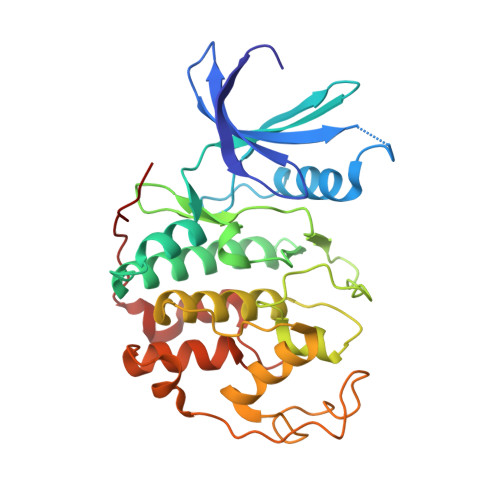
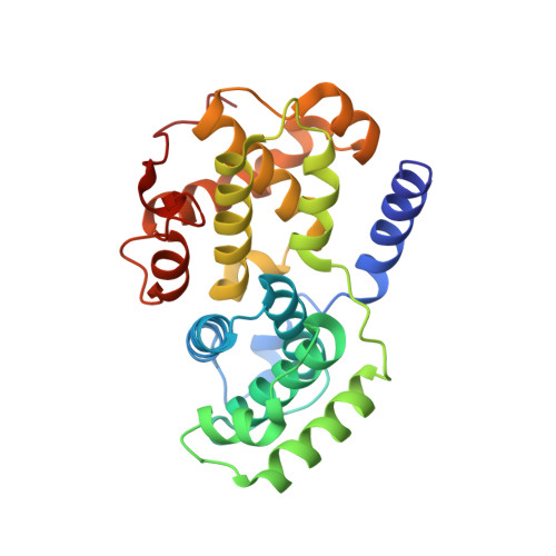
Synthesis and evaluation of pyrazolo[1,5-b]pyridazines as selective cyclin dependent kinase inhibitors.

Stevens, K.L.Reno, M.J.Alberti, J.B.Price, D.J.Kane-Carson, L.S.Knick, V.B.Shewchuk, L.M.Hassell, A.M.Veal, J.M.Davis, S.T.Griffin, R.J.Peel, M.R.

(2008) Bioorg Med Chem Lett 18: 5758-5762

  • DOI: https://doi.org/10.1016/j.bmcl.2008.09.069
  • Primary Citation of Related Structures:  
    3EID, 3EJ1

  • PubMed Abstract: 

    A novel series of pyrazolo[1,5-b]pyridazines have been synthesized and identified as cyclin dependant kinase inhibitors potentially useful for the treatment of solid tumors. Modification of the hinge-binding amine or the C(2)- and C(6)-substitutions on the pyrazolopyridazine core provided potent inhibitors of CDK4 and demonstrated enzyme selectivity against VEGFR-2 and GSK3beta.


  • Organizational Affiliation

    Department of Oncology, GlaxoSmithKline, Research Triangle Park, NC 27709, USA. Kirk.L.Stevens@gsk.com


Macromolecules
Find similar proteins by:  (by identity cutoff)  |  3D Structure
Entity ID: 1
MoleculeChains Sequence LengthOrganismDetailsImage
Cell division protein kinase 2
A, C
298Homo sapiensMutation(s): 0 
Gene Names: CDK2
EC: 2.7.11.22
UniProt & NIH Common Fund Data Resources
Find proteins for P24941 (Homo sapiens)
Explore P24941 
Go to UniProtKB:  P24941
PHAROS:  P24941
GTEx:  ENSG00000123374 
Entity Groups  
Sequence Clusters30% Identity50% Identity70% Identity90% Identity95% Identity100% Identity
UniProt GroupP24941
Sequence Annotations
Expand
  • Reference Sequence
Find similar proteins by:  (by identity cutoff)  |  3D Structure
Entity ID: 2
MoleculeChains Sequence LengthOrganismDetailsImage
Cyclin-A2
B, D
260Homo sapiensMutation(s): 0 
Gene Names: CCNA2CCN1CCNA
UniProt & NIH Common Fund Data Resources
Find proteins for P20248 (Homo sapiens)
Explore P20248 
Go to UniProtKB:  P20248
PHAROS:  P20248
GTEx:  ENSG00000145386 
Entity Groups  
Sequence Clusters30% Identity50% Identity70% Identity90% Identity95% Identity100% Identity
UniProt GroupP20248
Sequence Annotations
Expand
  • Reference Sequence
Small Molecules
Binding Affinity Annotations 
IDSourceBinding Affinity
5BP BindingDB:  3EJ1 IC50: 120 (nM) from 1 assay(s)
Experimental Data & Validation

Experimental Data

  • Method: X-RAY DIFFRACTION
  • Resolution: 3.22 Å
  • R-Value Free:  0.259 (Depositor), 0.260 (DCC) 
  • R-Value Work:  0.219 (Depositor), 0.220 (DCC) 
  • R-Value Observed: 0.221 (Depositor) 
Space Group: P 62 2 2
Unit Cell:
Length ( Å )Angle ( ˚ )
a = 183.776α = 90
b = 183.776β = 90
c = 214.045γ = 120
Software Package:
Software NamePurpose
REFMACrefinement
CNSrefinement
DENZOdata reduction
SCALEPACKdata scaling
CNSphasing

Structure Validation

View Full Validation Report


Ligand Structure Quality Assessment 

Created with Raphaël 2.3.0Worse 01 BetterLigand structure goodness of fit to experimental dataBest fitted 5BPClick on this verticalbar to view details

Entry History 

Deposition Data

Revision History  (Full details and data files)

  • Version 1.0: 2008-10-21
    Type: Initial release
  • Version 1.1: 2011-07-13
    Changes: Advisory, Refinement description, Version format compliance
  • Version 1.2: 2019-07-24
    Changes: Data collection, Refinement description
  • Version 1.3: 2023-08-30
    Changes: Data collection, Database references, Derived calculations, Refinement description