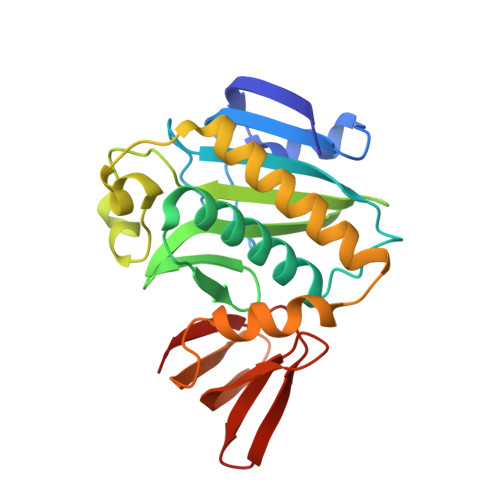
Structural and functional studies of the biotin protein ligase from Aquifex aeolicus reveal a critical role for a conserved residue in target specificity.
Tron, C.M., McNae, I.W., Nutley, M., Clarke, D.J., Cooper, A., Walkinshaw, M.D., Baxter, R.L., Campopiano, D.J.(2009) J Mol Biology 387: 129-146
- PubMed: 19385043 Search on PubMed
- DOI: https://doi.org/10.1016/j.jmb.2008.12.086
- Primary Citation Related Structures:
3EFR, 3EFS, 3FJP - PubMed Abstract:
Biotin protein ligase (BPL; EC 6.3.4.15) catalyses the formation of biotinyl-5'-AMP from biotin and ATP, and the succeeding biotinylation of the biotin carboxyl carrier protein. We describe the crystal structures, at 2.4 A resolution, of the class I BPL from the hyperthermophilic bacteria Aquifex aeolicus (AaBPL) in its ligand-free form and in complex with biotin and ATP. The solvent-exposed beta- and gamma-phosphates of ATP are located in the inter-subunit cavity formed by the N- and C-terminal domains. The Arg40 residue from the conserved GXGRXG motif is shown to interact with the carboxyl group of biotin and to stabilise the alpha- and beta-phosphates of the nucleotide. The structure of the mutant AaBPL R40G in both the ligand-free and biotin-bound forms reveals that the mutated loop has collapsed, thus hindering ATP binding. Isothermal titration calorimetry indicated that the presence of biotin is not required for ATP binding to wild-type AaBPL in the absence of Mg(2+), and the binding of biotin and ATP has been determined to occur via a random but cooperative process. The affinity for biotin is relatively unaffected by the R40G mutation. In contrast, the thermodynamic data indicate that binding of ATP to AaBPL R40G is very weak in the absence or in the presence of biotin. The AaBPL R40G mutant remains catalytically active but shows poor substrate specificity; mass spectrometry and Western blot studies revealed that the mutant biotinylates both the target A. aeolicus BCCPDelta67 fragment and BSA, and is subject to self-biotinylation.
- School of Chemistry, EaStCHEM, The University of Edinburgh, West Mains Road, King's Buildings, Edinburgh, Scotland, UK.
Organizational Affiliation: