


High resolution crystal structure of the organic hydroperoxide resistance protein from Vibrio cholerae O1 biovar eltor str. N16961
Nocek, B., Mulligan, R., Kwon, K., Anderson, W.F., Joachimiak, A.To be published.
Experimental Data Snapshot
wwPDB Validation 3D Report Full Report

Entity ID: 1 | |||||
|---|---|---|---|---|---|
| Molecule | Chains | Sequence Length | Organism | Details | Image |
| Organic hydroperoxide resistance protein, putative | 148 | Vibrio cholerae O1 biovar El Tor str. N16961 | Mutation(s): 0 Gene Names: VC_A1006 | 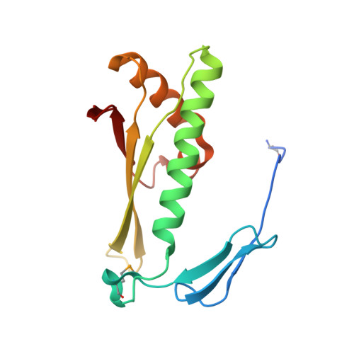 | |
UniProt | |||||
Entity Groups | |||||
| Sequence Clusters | 30% Identity50% Identity70% Identity90% Identity95% Identity100% Identity | ||||
| UniProt Group | Q9KKU4 | ||||
Sequence AnnotationsExpand | |||||
Reference Sequence | |||||
| Ligands 3 Unique | |||||
|---|---|---|---|---|---|
| ID | Chains | Name / Formula / InChI Key | 2D Diagram | 3D Interactions | |
| IMD Download:Ideal Coordinates CCD File | O [auth A] | IMIDAZOLE C3 H5 N2 RAXXELZNTBOGNW-UHFFFAOYSA-O |  | ||
| ZN Download:Ideal Coordinates CCD File | I [auth A] J [auth A] K [auth A] L [auth A] M [auth A] | ZINC ION Zn PTFCDOFLOPIGGS-UHFFFAOYSA-N |  | ||
| ACT Download:Ideal Coordinates CCD File | B [auth A] C [auth A] D [auth A] E [auth A] F [auth A] | ACETATE ION C2 H3 O2 QTBSBXVTEAMEQO-UHFFFAOYSA-M |  | ||
| Modified Residues 1 Unique | |||||
|---|---|---|---|---|---|
| ID | Chains | Type | Formula | 2D Diagram | Parent |
| MSE Query on MSE | A | L-PEPTIDE LINKING | C5 H11 N O2 Se |  | MET |
| Length ( Å ) | Angle ( ˚ ) |
|---|---|
| a = 59.649 | α = 90 |
| b = 71.127 | β = 90 |
| c = 71.629 | γ = 90 |
| Software Name | Purpose |
|---|---|
| SBC-Collect | data collection |
| MOLREP | phasing |
| CCP4 | model building |
| REFMAC | refinement |
| HKL-3000 | data reduction |
| HKL-3000 | data scaling |
| HKL-3000 | phasing |
| DM | phasing |
| ARP/wARP | model building |
| CCP4 | phasing |
| Coot | model building |


