3EEE | pdb_00003eee

Probing the function of heme distortion in the H-NOX family


Experimental Data Snapshot

  • Method: X-RAY DIFFRACTION
  • Resolution: 2.12 Å
  • R-Value Free: 
    0.252 (Depositor), 0.248 (DCC) 
  • R-Value Work: 
    0.205 (Depositor), 0.202 (DCC) 
  • R-Value Observed: 
    0.207 (Depositor) 

Starting Model: experimental
View more details

wwPDB Validation 3D Report Full Report

Validation slider image for 3EEE

Ligand Structure Quality Assessment 


This is version 1.3 of the entry. See complete history

Literature

Probing the function of heme distortion in the H-NOX family.

Olea, C.Boon, E.M.Pellicena, P.Kuriyan, J.Marletta, M.A.

(2008) ACS Chem Biol 3: 703-710

  • DOI: https://doi.org/10.1021/cb800185h
  • Primary Citation Related Structures: 
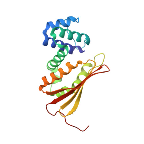
    3EEE

  • PubMed Abstract: 

    Hemoproteins carry out diverse functions utilizing a wide range of chemical reactivity while employing the same heme prosthetic group. It is clear from high-resolution crystal structures and biochemical studies that protein-bound hemes are not planar and adopt diverse conformations. The crystal structure of an H-NOX domain from Thermoanaerobacter tengcongensis (Tt H-NOX) contains the most distorted heme reported to date. In this study, Tt H-NOX was engineered to adopt a flatter heme by mutating proline 115, a conserved residue in the H-NOX family, to alanine. Decreasing heme distortion in Tt H-NOX increases affinity for oxygen and decreases the reduction potential of the heme iron. Additionally, flattening the heme is associated with significant shifts in the N-terminus of the protein. These results show a clear link between the heme conformation and Tt H-NOX structure and demonstrate that heme distortion is an important determinant for maintaining biochemical properties in H-NOX proteins.


  • Organizational Affiliation
    • Department of Molecular and Cell Biology, California Institute for Quantitative Biosciences, University of California, Berkeley, California 94720, USA.

Macromolecule Content 

  • Total Structure Weight: 91.04 kDa 
  • Atom Count: 6,557 
  • Modeled Residue Count: 752 
  • Deposited Residue Count: 752 
  • Unique protein chains: 1

Macromolecules

Find similar proteins by:|  3D Structure
Entity ID: 1
MoleculeChains  Sequence LengthOrganismDetailsImage
Methyl-accepting chemotaxis protein
A, B, C, D
188Caldanaerobacter subterraneus subsp. tengcongensisMutation(s): 1 
Gene Names: Tar4
Membrane Entity: Yes 
UniProt
Find proteins for Q8RBX6 (Caldanaerobacter subterraneus subsp. tengcongensis (strain DSM 15242 / JCM 11007 / NBRC 100824 / MB4))
Explore Q8RBX6 
Go to UniProtKB:  Q8RBX6
Entity Groups
Sequence Clusters30% Identity50% Identity70% Identity90% Identity95% Identity100% Identity
UniProt GroupQ8RBX6
Sequence Annotations
Expand
Reference Sequence

Small Molecules

Ligands 4 Unique
IDChains Name / Formula / InChI Key2D Diagram3D Interactions
HEM

Query on HEM


Download:Ideal Coordinates CCD File
E [auth A],
H [auth B],
L [auth C],
O [auth D]
PROTOPORPHYRIN IX CONTAINING FE
C34 H32 Fe N4 O4
KABFMIBPWCXCRK-RGGAHWMASA-L
SO4

Query on SO4


Download:Ideal Coordinates CCD File
G [auth A],
K [auth B],
Q [auth D]
SULFATE ION
O4 S
QAOWNCQODCNURD-UHFFFAOYSA-L
CL

Query on CL


Download:Ideal Coordinates CCD File
J [auth B],
N [auth C]
CHLORIDE ION
Cl
VEXZGXHMUGYJMC-UHFFFAOYSA-M
OXY

Query on OXY


Download:Ideal Coordinates CCD File
F [auth A],
I [auth B],
M [auth C],
P [auth D]
OXYGEN MOLECULE
O2
MYMOFIZGZYHOMD-UHFFFAOYSA-N

Experimental Data & Validation

Experimental Data

  • Method: X-RAY DIFFRACTION
  • Resolution: 2.12 Å
  • R-Value Free:  0.252 (Depositor), 0.248 (DCC) 
  • R-Value Work:  0.205 (Depositor), 0.202 (DCC) 
  • R-Value Observed: 0.207 (Depositor) 
Space Group: C 1 2 1
Unit Cell:
Length ( Å )Angle ( ˚ )
a = 116.253α = 90
b = 124.686β = 126.58
c = 83.555γ = 90
Software Package:
Software NamePurpose
PHENIXrefinement
PDB_EXTRACTdata extraction
HKL-2000data collection
HKL-2000data reduction
HKL-2000data scaling
PHASERphasing

Structure Validation

View Full Validation Report


Ligand Structure Quality Assessment 


Entry History 

Deposition Data

Revision History  (Full details and data files)

  • Version 1.0: 2008-11-25
    Type: Initial release
  • Version 1.1: 2011-07-13
    Changes: Version format compliance
  • Version 1.2: 2021-10-20
    Changes: Database references, Derived calculations
  • Version 1.3: 2023-08-30
    Changes: Data collection, Refinement description