3EB8 | pdb_00003eb8

VirA


Experimental Data Snapshot

  • Method: X-RAY DIFFRACTION
  • Resolution: 2.40 Å
  • R-Value Free: 
    0.267 (Depositor) 
  • R-Value Work: 
    0.217 (Depositor), 0.216 (DCC) 
  • R-Value Observed: 
    0.220 (Depositor) 

wwPDB Validation 3D Report Full Report

Validation slider image for 3EB8

This is version 1.3 of the entry. See complete history

Literature

Structural and functional studies indicate that Shigella VirA is not a protease and does not directly destabilize microtubules.

Germane, K.L.Ohi, R.Goldberg, M.B.Spiller, B.W.

(2008) Biochemistry 47: 10241-10243

  • DOI: https://doi.org/10.1021/bi801533k
  • Primary Citation Related Structures: 
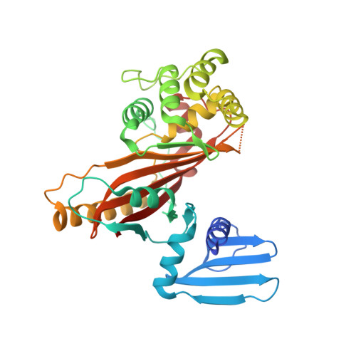
    3EB8

  • PubMed Abstract: 

    VirA, an essential virulence factor in Shigella disease pathogenesis, is involved in the uptake, motility, and cell-to-cell spread of Shigella organisms within the human host. These functions have been attributed to a VirA protease activity and a mechanism of microtubule destruction via tubulin degradation [Yoshida, S., et al. (2006) Science 314, 985-989]. We report functional and crystallographic data indicating a novel VirA structure that lacks these activities but highlights the homology to the EspG virulence factor of pathogenic Escherichia coli.


  • Organizational Affiliation
    • Department of Pharmacology, Vanderbilt University Medical Center, 461 Preston Research Building, 2220 Pierce Avenue, Nashville, Tennessee 37232, USA.

Macromolecule Content 

  • Total Structure Weight: 79.75 kDa 
  • Atom Count: 5,371 
  • Modeled Residue Count: 670 
  • Deposited Residue Count: 716 
  • Unique protein chains: 1

Macromolecules

Find similar proteins by:|  3D Structure
Entity ID: 1
MoleculeChains  Sequence LengthOrganismDetailsImage
Cysteine protease-like virA
A, B
358Shigella flexneriMutation(s): 0 
Gene Names: virACP0181pWR501_0191
EC: 3.4.22
UniProt
Find proteins for Q7BU69 (Shigella flexneri)
Explore Q7BU69 
Go to UniProtKB:  Q7BU69
Entity Groups
Sequence Clusters30% Identity50% Identity70% Identity90% Identity95% Identity100% Identity
UniProt GroupQ7BU69
Sequence Annotations
Expand
Reference Sequence

Experimental Data & Validation

Experimental Data

  • Method: X-RAY DIFFRACTION
  • Resolution: 2.40 Å
  • R-Value Free:  0.267 (Depositor) 
  • R-Value Work:  0.217 (Depositor), 0.216 (DCC) 
  • R-Value Observed: 0.220 (Depositor) 
Space Group: P 41 21 2
Unit Cell:
Length ( Å )Angle ( ˚ )
a = 91.29α = 90
b = 91.29β = 90
c = 198.29γ = 90
Software Package:
Software NamePurpose
ADSCdata collection
SHARPphasing
PHENIXrefinement
HKL-2000data reduction
HKL-2000data scaling

Structure Validation

View Full Validation Report


Entry History 

Deposition Data

Revision History  (Full details and data files)

  • Version 1.0: 2008-09-30
    Type: Initial release
  • Version 1.1: 2011-07-13
    Changes: Derived calculations, Version format compliance
  • Version 1.2: 2020-02-26
    Changes: Data collection, Database references
  • Version 1.3: 2024-02-21
    Changes: Data collection, Database references