


Crystal structure of Schistosoma mansoni purine nucleoside phosphorylase (SmPNP) in complex with adenine, 8-aminoguanine, 8-azaguanine and 6-chloroguanine.
Pereira, H.M., Rezende, M.M., Oliva, G., Garratt, R.C.To be published.
Experimental Data Snapshot
Starting Model: experimental
View more details
Entity ID: 1 | |||||
|---|---|---|---|---|---|
| Molecule | Chains | Sequence Length | Organism | Details | Image |
| Purine-nucleoside phosphorylase | 287 | Schistosoma mansoni | Mutation(s): 0 Gene Names: SmPNP EC: 2.4.2.1 | 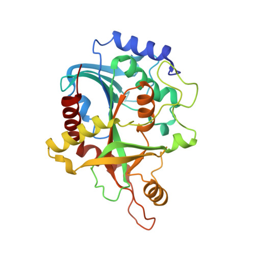 | |
UniProt | |||||
Entity Groups | |||||
| Sequence Clusters | 30% Identity50% Identity70% Identity90% Identity95% Identity100% Identity | ||||
| UniProt Group | Q9BMI9 | ||||
Sequence AnnotationsExpand | |||||
Reference Sequence | |||||
| Ligands 4 Unique | |||||
|---|---|---|---|---|---|
| ID | Chains | Name / Formula / InChI Key | 2D Diagram | 3D Interactions | |
| 6GU Download:Ideal Coordinates CCD File | F [auth A], L [auth C] | 6-chloroguanine C5 H4 Cl N5 RYYIULNRIVUMTQ-UHFFFAOYSA-N |  | ||
| SO4 Download:Ideal Coordinates CCD File | E [auth A], J [auth B], M [auth C] | SULFATE ION O4 S QAOWNCQODCNURD-UHFFFAOYSA-L |  | ||
| DMS Download:Ideal Coordinates CCD File | D [auth A], G [auth A], H [auth B], K [auth C] | DIMETHYL SULFOXIDE C2 H6 O S IAZDPXIOMUYVGZ-UHFFFAOYSA-N |  | ||
| ACT Download:Ideal Coordinates CCD File | I [auth B] | ACETATE ION C2 H3 O2 QTBSBXVTEAMEQO-UHFFFAOYSA-M |  | ||
| Length ( Å ) | Angle ( ˚ ) |
|---|---|
| a = 46.821 | α = 90 |
| b = 118.121 | β = 90 |
| c = 129.055 | γ = 90 |
| Software Name | Purpose |
|---|---|
| MOSFLM | data reduction |
| SCALA | data scaling |
| PHENIX | refinement |
| PDB_EXTRACT | data extraction |
| MAR345dtb | data collection |
| REFMAC | phasing |


