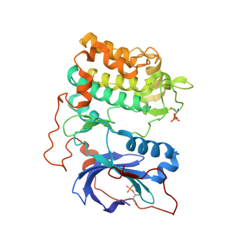
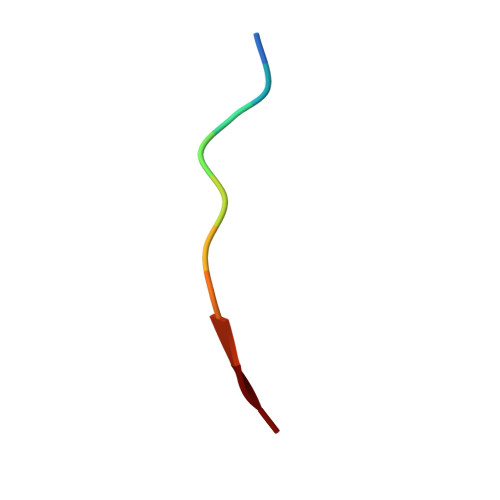
3E87

Crystal structures of the kinase domain of AKT2 in complex with ATP-competitive inhibitors


Experimental Data Snapshot

  • Method: X-RAY DIFFRACTION
  • Resolution: 2.30 Å
  • R-Value Free: 0.246 
  • R-Value Work: 0.225 
  • R-Value Observed: 0.226 

wwPDB Validation   3D Report Full Report


Ligand Structure Quality Assessment 


This is version 1.4 of the entry. See complete history


Literature

Aminofurazans as potent inhibitors of AKT kinase

Rouse, M.B.Seefeld, M.A.Leber, J.D.McNulty, K.C.Sun, L.Miller, W.H.Zhang, S.Minthorn, E.A.Concha, N.O.Choudhry, A.E.Schaber, M.D.Heerding, D.A.

(2009) Bioorg Med Chem Lett 19: 1508-1511

  • DOI: https://doi.org/10.1016/j.bmcl.2009.01.002
  • Primary Citation of Related Structures:  
    3E87, 3E88, 3E8C, 3E8D, 3E8E

  • PubMed Abstract: 

    AKT inhibitors containing an imidazopyridine aminofurazan scaffold have been optimized. We have previously disclosed identification of the AKT inhibitor GSK690693, which has been evaluated in clinical trials in cancer patients. Herein we describe recent efforts focusing on investigating a distinct region of this scaffold that have afforded compounds (30 and 32) with comparable activity profiles to that of GSK690693.


  • Organizational Affiliation

    Oncology Chemistry, GlaxoSmithKline, Collegeville, PA 19426, USA.


Macromolecules
Find similar proteins by:  (by identity cutoff)  |  3D Structure
Entity ID: 1
MoleculeChains Sequence LengthOrganismDetailsImage
RAC-beta serine/threonine-protein kinase
A, B
335Homo sapiensMutation(s): 1 
Gene Names: AKT2
EC: 2.7.11.1
UniProt & NIH Common Fund Data Resources
Find proteins for P31751 (Homo sapiens)
Explore P31751 
Go to UniProtKB:  P31751
PHAROS:  P31751
GTEx:  ENSG00000105221 
Entity Groups  
Sequence Clusters30% Identity50% Identity70% Identity90% Identity95% Identity100% Identity
UniProt GroupP31751
Sequence Annotations
Expand
  • Reference Sequence

Find similar proteins by:  Sequence   |   3D Structure  

Entity ID: 2
MoleculeChains Sequence LengthOrganismDetailsImage
Glycogen synthase kinase-3 beta peptide
C, D
10N/AMutation(s): 0 
EC: 2.7.11.1 (UniProt), 2.7.11.26 (UniProt)
UniProt & NIH Common Fund Data Resources
Find proteins for P49841 (Homo sapiens)
Explore P49841 
Go to UniProtKB:  P49841
PHAROS:  P49841
GTEx:  ENSG00000082701 
Entity Groups  
Sequence Clusters30% Identity50% Identity70% Identity90% Identity95% Identity100% Identity
UniProt GroupP49841
Sequence Annotations
Expand
  • Reference Sequence
Small Molecules
Ligands 1 Unique
IDChains Name / Formula / InChI Key2D Diagram3D Interactions
G95
Query on G95

Download Ideal Coordinates CCD File 
E [auth A],
F [auth B]
N-[(1S)-2-amino-1-phenylethyl]-5-(1H-pyrrolo[2,3-b]pyridin-4-yl)thiophene-2-carboxamide
C20 H18 N4 O S
TWYNGDRSMHRPSY-MRXNPFEDSA-N
Modified Residues  1 Unique
IDChains TypeFormula2D DiagramParent
TPO
Query on TPO
A, B
L-PEPTIDE LINKINGC4 H10 N O6 PTHR
Binding Affinity Annotations 
IDSourceBinding Affinity
G95 BindingDB:  3E87 IC50: min: 6, max: 190 (nM) from 2 assay(s)
Experimental Data & Validation

Experimental Data

  • Method: X-RAY DIFFRACTION
  • Resolution: 2.30 Å
  • R-Value Free: 0.246 
  • R-Value Work: 0.225 
  • R-Value Observed: 0.226 
  • Space Group: P 32
Unit Cell:
Length ( Å )Angle ( ˚ )
a = 117.323α = 90
b = 117.323β = 90
c = 45.045γ = 120
Software Package:
Software NamePurpose
DENZOdata reduction
SCALEPACKdata scaling
REFMACrefinement
PDB_EXTRACTdata extraction
JDirectordata collection
HKL-2000data reduction
PHASERphasing

Structure Validation

View Full Validation Report


Ligand Structure Quality Assessment 


Entry History 

Deposition Data

Revision History  (Full details and data files)

  • Version 1.0: 2008-10-14
    Type: Initial release
  • Version 1.1: 2011-07-13
    Changes: Advisory, Version format compliance
  • Version 1.2: 2017-10-25
    Changes: Refinement description
  • Version 1.3: 2021-10-20
    Changes: Database references, Derived calculations
  • Version 1.4: 2024-11-20
    Changes: Data collection, Refinement description, Structure summary