3E76 | pdb_00003e76

Crystal structure of Wild-type GroEL with bound Thallium ions


Experimental Data Snapshot

  • Method: X-RAY DIFFRACTION
  • Resolution: 3.94 Å
  • R-Value Free: 
    0.293 (Depositor), 0.277 (DCC) 
  • R-Value Work: 
    0.261 (Depositor), 0.249 (DCC) 
  • R-Value Observed: 
    0.262 (Depositor) 

Starting Model: experimental
View more details

wwPDB Validation 3D Report Full Report

Validation slider image for 3E76

Ligand Structure Quality Assessment 


This is version 1.2 of the entry. See complete history

Literature

Use of thallium to identify monovalent cation binding sites in GroEL.

Kiser, P.D.Lorimer, G.H.Palczewski, K.

(2009) Acta Crystallogr Sect F Struct Biol Cryst Commun 65: 967-971

  • DOI: https://doi.org/10.1107/S1744309109032928
  • Primary Citation Related Structures: 
    3E76

  • PubMed Abstract: 

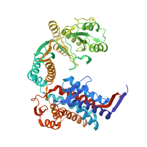
    GroEL is a bacterial chaperone protein that assembles into a homotetradecameric complex exhibiting D(7) symmetry and utilizes the co-chaperone protein GroES and ATP hydrolysis to assist in the proper folding of a variety of cytosolic proteins. GroEL utilizes two metal cofactors, Mg(2+) and K(+), to bind and hydrolyze ATP. A K(+)-binding site has been proposed to be located next to the nucleotide-binding site, but the available structural data do not firmly support this conclusion. Moreover, more than one functionally significant K(+)-binding site may exist within GroEL. Because K(+) has important and complex effects on GroEL activity and is involved in both positive (intra-ring) and negative (inter-ring) cooperativity for ATP hydrolysis, it is important to determine the exact location of these cation-binding site(s) within GroEL. In this study, the K(+) mimetic Tl(+) was incorporated into GroEL crystals, a moderately redundant 3.94 A resolution X-ray diffraction data set was collected from a single crystal and the strong anomalous scattering signal from the thallium ion was used to identify monovalent cation-binding sites. The results confirmed the previously proposed placement of K(+) next to the nucleotide-binding site and also identified additional binding sites that may be important for GroEL function and cooperativity. These findings also demonstrate the general usefulness of Tl(+) for the identification of monovalent cation-binding sites in protein crystal structures, even when the quality and resolution of the diffraction data are relatively low.


  • Organizational Affiliation
    • Case Western Reserve University, USA.

Macromolecule Content 

  • Total Structure Weight: 818.71 kDa 
  • Atom Count: 54,464 
  • Modeled Residue Count: 7,336 
  • Deposited Residue Count: 7,658 
  • Unique protein chains: 1

Macromolecules

Find similar proteins by:|  3D Structure
Entity ID: 1
MoleculeChains  Sequence LengthOrganismDetailsImage
60 kDa chaperonin
A, B, C, D, E
A, B, C, D, E, F, G, H, I, J, K, L, M, N
547Escherichia coli UTI89Mutation(s): 0 
Gene Names: groLgroELUTI89_C4741
EC: 3.6.4.9 (PDB Primary Data), 5.6.1.7 (UniProt)
UniProt
Find proteins for Q1R3B6 (Escherichia coli (strain UTI89 / UPEC))
Explore Q1R3B6 
Go to UniProtKB:  Q1R3B6
Entity Groups
Sequence Clusters30% Identity50% Identity70% Identity90% Identity95% Identity100% Identity
UniProt GroupQ1R3B6
Sequence Annotations
Expand
Reference Sequence

Small Molecules

Ligands 3 Unique
IDChains Name / Formula / InChI Key2D Diagram3D Interactions
AGS

Query on AGS


Download:Ideal Coordinates CCD File
AA [auth C]
BB [auth H]
BC [auth M]
FA [auth D]
GC [auth N]
AA [auth C],
BB [auth H],
BC [auth M],
FA [auth D],
GC [auth N],
HB [auth I],
LA [auth E],
MB [auth J],
O [auth A],
QA [auth F],
RB [auth K],
U [auth B],
WA [auth G],
WB [auth L]
PHOSPHOTHIOPHOSPHORIC ACID-ADENYLATE ESTER
C10 H16 N5 O12 P3 S
NLTUCYMLOPLUHL-KQYNXXCUSA-N
TL

Query on TL


Download:Ideal Coordinates CCD File
BA [auth C]
CA [auth C]
CB [auth H]
CC [auth M]
DA [auth C]
BA [auth C],
CA [auth C],
CB [auth H],
CC [auth M],
DA [auth C],
DB [auth H],
DC [auth M],
EB [auth H],
EC [auth M],
GA [auth D],
GB [auth H],
HA [auth D],
HC [auth N],
IA [auth D],
IB [auth I],
IC [auth N],
JB [auth I],
KA [auth D],
KB [auth I],
MA [auth E],
NA [auth E],
NB [auth J],
OA [auth E],
OB [auth J],
P [auth A],
PB [auth J],
Q [auth A],
R [auth A],
RA [auth F],
SA [auth F],
SB [auth K],
T [auth A],
TA [auth F],
TB [auth K],
UB [auth K],
V [auth B],
VA [auth F],
W [auth B],
X [auth B],
XA [auth G],
XB [auth L],
YA [auth G],
YB [auth L],
Z [auth B],
ZA [auth G],
ZB [auth L]
THALLIUM (I) ION
Tl
ZLUSCZLCHQSJRU-UHFFFAOYSA-N
MG

Query on MG


Download:Ideal Coordinates CCD File
AB [auth G]
AC [auth L]
EA [auth C]
FB [auth H]
FC [auth M]
AB [auth G],
AC [auth L],
EA [auth C],
FB [auth H],
FC [auth M],
JA [auth D],
JC [auth N],
LB [auth I],
PA [auth E],
QB [auth J],
S [auth A],
UA [auth F],
VB [auth K],
Y [auth B]
MAGNESIUM ION
Mg
JLVVSXFLKOJNIY-UHFFFAOYSA-N

Experimental Data & Validation

Experimental Data

  • Method: X-RAY DIFFRACTION
  • Resolution: 3.94 Å
  • R-Value Free:  0.293 (Depositor), 0.277 (DCC) 
  • R-Value Work:  0.261 (Depositor), 0.249 (DCC) 
  • R-Value Observed: 0.262 (Depositor) 
Space Group: P 21 21 21
Unit Cell:
Length ( Å )Angle ( ˚ )
a = 135.676α = 90
b = 260.954β = 90
c = 287.872γ = 90
Software Package:
Software NamePurpose
REFMACrefinement
HKL-2000data collection
HKL-2000data reduction
HKL-2000data scaling
PHASERphasing

Structure Validation

View Full Validation Report


Ligand Structure Quality Assessment 


Entry History 

Deposition Data

Revision History  (Full details and data files)

  • Version 1.0: 2009-08-25
    Type: Initial release
  • Version 1.1: 2011-07-13
    Changes: Advisory, Version format compliance
  • Version 1.2: 2023-08-30
    Changes: Data collection, Database references, Derived calculations, Refinement description